

A novel abnormality annotation database for COVID-19 affected frontal lung X-rays
- PMID: 36240175
- PMCID: PMC9565456
- DOI: 10.1371/journal.pone.0271931
A novel abnormality annotation database for COVID-19 affected frontal lung X-rays
Abstract
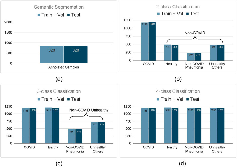
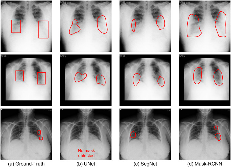
Consistent clinical observations of characteristic findings of COVID-19 pneumonia on chest X-rays have attracted the research community to strive to provide a fast and reliable method for screening suspected patients. Several machine learning algorithms have been proposed to find the abnormalities in the lungs using chest X-rays specific to COVID-19 pneumonia and distinguish them from other etiologies of pneumonia. However, despite the enormous magnitude of the pandemic, there are very few instances of public databases of COVID-19 pneumonia, and to the best of our knowledge, there is no database with annotation of abnormalities on the chest X-rays of COVID-19 affected patients. Annotated databases of X-rays can be of significant value in the design and development of algorithms for disease prediction. Further, explainability analysis for the performance of existing or new deep learning algorithms will be enhanced significantly with access to ground-truth abnormality annotations. The proposed COVID Abnormality Annotation for X-Rays (CAAXR) database is built upon the BIMCV-COVID19+ database which is a large-scale dataset containing COVID-19+ chest X-rays. The primary contribution of this study is the annotation of the abnormalities in over 1700 frontal chest X-rays. Further, we define protocols for semantic segmentation as well as classification for robust evaluation of algorithms. We provide benchmark results on the defined protocols using popular deep learning models such as DenseNet, ResNet, MobileNet, and VGG for classification, and UNet, SegNet, and Mask-RCNN for semantic segmentation. The classwise accuracy, sensitivity, and AUC-ROC scores are reported for the classification models, and the IoU and DICE scores are reported for the segmentation models.
Conflict of interest statement
The authors have declared that no competing interests exist.
Figures
Similar articles
-
A radiomics-boosted deep-learning model for COVID-19 and non-COVID-19 pneumonia classification using chest x-ray images.Med Phys. 2022 May;49(5):3213-3222. doi: 10.1002/mp.15582. Epub 2022 Mar 15. Med Phys. 2022. PMID: 35263458 Free PMC article.
-
DenResCov-19: A deep transfer learning network for robust automatic classification of COVID-19, pneumonia, and tuberculosis from X-rays.Comput Med Imaging Graph. 2021 Dec;94:102008. doi: 10.1016/j.compmedimag.2021.102008. Epub 2021 Oct 23. Comput Med Imaging Graph. 2021. PMID: 34763146 Free PMC article.
-
COVID-19 infection localization and severity grading from chest X-ray images.Comput Biol Med. 2021 Dec;139:105002. doi: 10.1016/j.compbiomed.2021.105002. Epub 2021 Oct 30. Comput Biol Med. 2021. PMID: 34749094 Free PMC article.
-
StackNet-DenVIS: a multi-layer perceptron stacked ensembling approach for COVID-19 detection using X-ray images.Phys Eng Sci Med. 2020 Dec;43(4):1399-1414. doi: 10.1007/s13246-020-00952-6. Epub 2020 Dec 4. Phys Eng Sci Med. 2020. PMID: 33275187 Free PMC article.
-
COVID Detection From Chest X-Ray Images Using Multi-Scale Attention.IEEE J Biomed Health Inform. 2022 Apr;26(4):1496-1505. doi: 10.1109/JBHI.2022.3151171. Epub 2022 Apr 14. IEEE J Biomed Health Inform. 2022. PMID: 35157603
Cited by
-
Automated prediction of COVID-19 severity upon admission by chest X-ray images and clinical metadata aiming at accuracy and explainability.Sci Rep. 2023 Mar 14;13(1):4226. doi: 10.1038/s41598-023-30505-2. Sci Rep. 2023. PMID: 36918593 Free PMC article.
-
FedSGDCOVID: Federated SGD COVID-19 Detection under Local Differential Privacy Using Chest X-ray Images and Symptom Information.Sensors (Basel). 2022 May 13;22(10):3728. doi: 10.3390/s22103728. Sensors (Basel). 2022. PMID: 35632136 Free PMC article.
-
Frequency of Missed Findings on Chest Radiographs (CXRs) in an International, Multicenter Study: Application of AI to Reduce Missed Findings.Diagnostics (Basel). 2022 Sep 30;12(10):2382. doi: 10.3390/diagnostics12102382. Diagnostics (Basel). 2022. PMID: 36292071 Free PMC article.
-
AI-based radiodiagnosis using chest X-rays: A review.Front Big Data. 2023 Apr 6;6:1120989. doi: 10.3389/fdata.2023.1120989. eCollection 2023. Front Big Data. 2023. PMID: 37091458 Free PMC article. Review.
-
Public Covid-19 X-ray datasets and their impact on model bias - A systematic review of a significant problem.Med Image Anal. 2021 Dec;74:102225. doi: 10.1016/j.media.2021.102225. Epub 2021 Sep 28. Med Image Anal. 2021. PMID: 34597937 Free PMC article.
References
-
- Schiffmann A. World COVID-19 Stats; 2020. https://ncov2019.live/.
-
- Venugopal VK, Mahajan V, Rajan S, Agarwal VK, Rajan R, Syed S, et al.. A Systematic Meta-Analysis of CT Features of COVID-19: Lessons from Radiology. medRxiv. 2020;.
-
- Rodriguez-Morales AJ, Cardona-Ospina JA, Gutiérrez-Ocampo E, Villamizar-Peña R, Holguin-Rivera Y, Escalera-Antezana JP, et al.. Clinical, laboratory and imaging features of COVID-19: A systematic review and meta-analysis. Travel medicine and infectious disease. 2020; p. 101623. doi: 10.1016/j.tmaid.2020.101623 - DOI - PMC - PubMed
Publication types
MeSH terms
LinkOut - more resources
Full Text Sources
Medical
