. 2022 Oct 14;18(10):e1010448.
doi: 10.1371/journal.pgen.1010448. eCollection 2022 Oct.

Nascent polypeptide-Associated Complex and Signal Recognition Particle have cardiac-specific roles in heart development and remodeling

Affiliations

Nascent polypeptide-Associated Complex and Signal Recognition Particle have cardiac-specific roles in heart development and remodeling

Analyne M Schroeder et al. PLoS Genet. .

Abstract

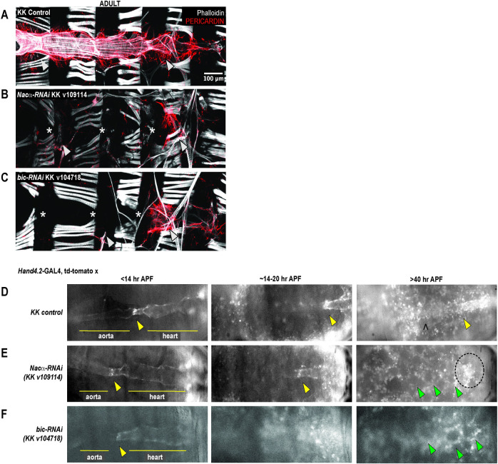
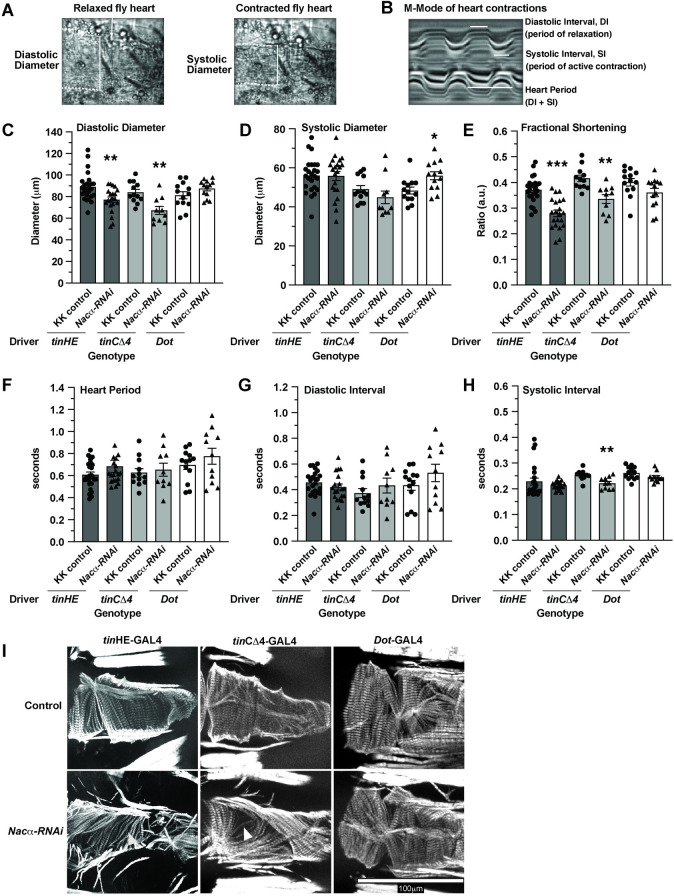
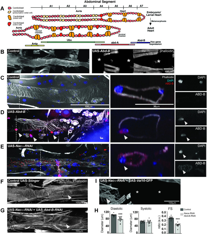
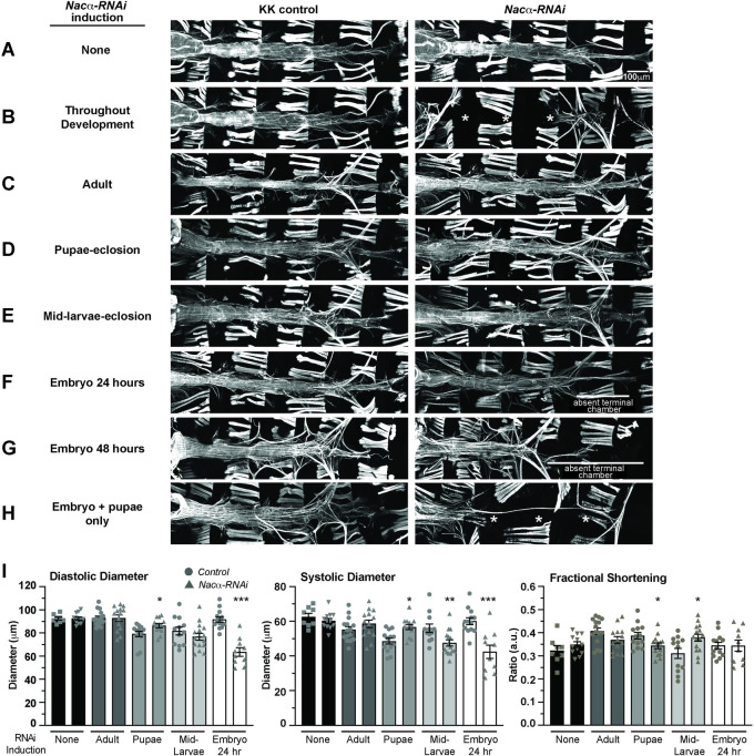
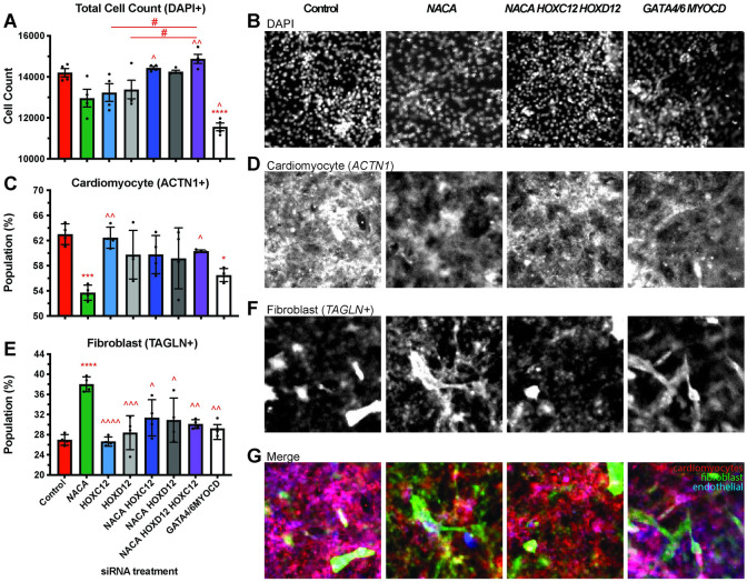
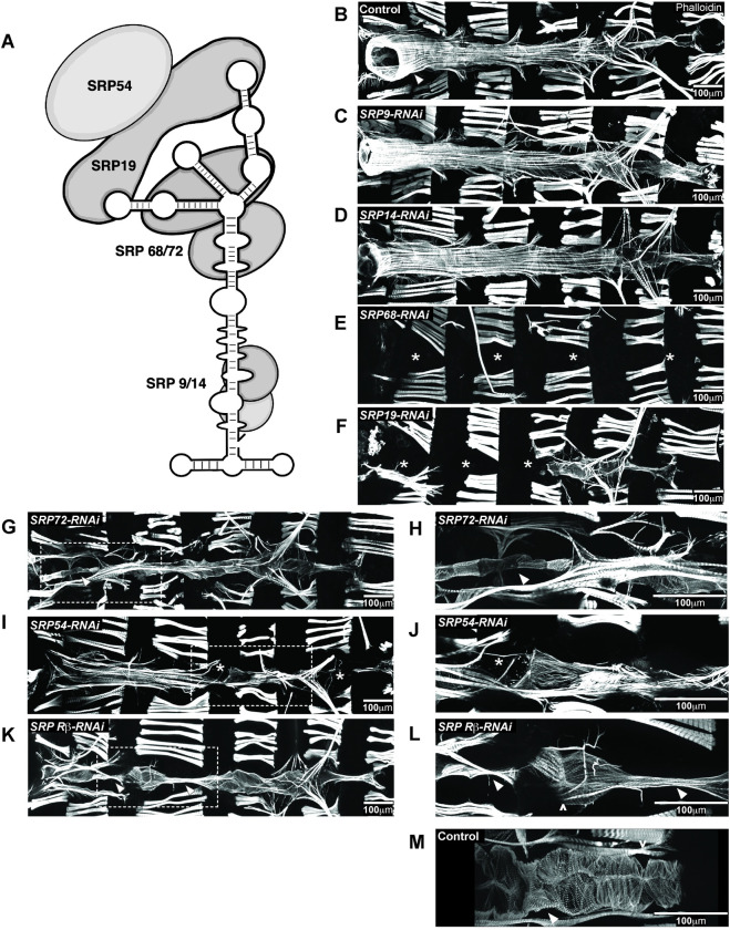
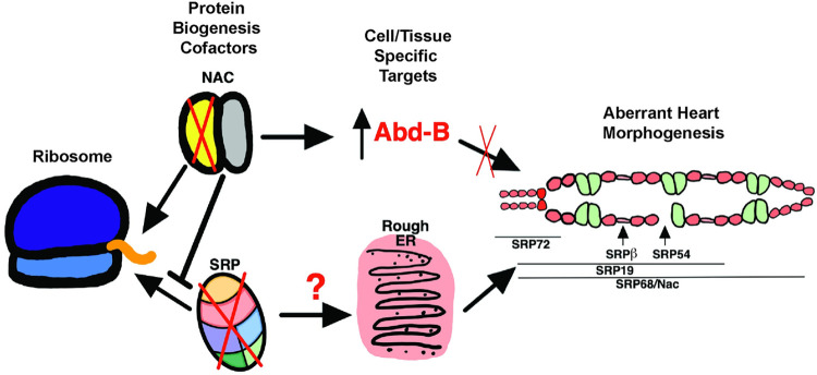
Establishing a catalog of Congenital Heart Disease (CHD) genes and identifying functional networks would improve our understanding of its oligogenic underpinnings. Our studies identified protein biogenesis cofactors Nascent polypeptide-Associated Complex (NAC) and Signal-Recognition-Particle (SRP) as disease candidates and novel regulators of cardiac differentiation and morphogenesis. Knockdown (KD) of the alpha- (Nacα) or beta-subunit (bicaudal, bic) of NAC in the developing Drosophila heart disrupted cardiac developmental remodeling resulting in a fly with no heart. Heart loss was rescued by combined KD of Nacα with the posterior patterning Hox gene Abd-B. Consistent with a central role for this interaction in cardiogenesis, KD of Nacα in cardiac progenitors derived from human iPSCs impaired cardiac differentiation while co-KD with human HOXC12 and HOXD12 rescued this phenotype. Our data suggest that Nacα KD preprograms cardioblasts in the embryo for abortive remodeling later during metamorphosis, as Nacα KD during translation-intensive larval growth or pupal remodeling only causes moderate heart defects. KD of SRP subunits in the developing fly heart produced phenotypes that targeted specific segments and cell types, again suggesting cardiac-specific and spatially regulated activities. Together, we demonstrated directed function for NAC and SRP in heart development, and that regulation of NAC function depends on Hox genes.


Conflict of interest statement

The authors have declared that no competing interests exist.

Figures

Fig 1
Fig 1. Knockdown (KD) of either subunit of Nascent polypeptide Associated Complex led to complete histolysis of the heart in adult flies.
Hand4.2-GAL4 driving heart specific expression of A, KK control B, Nacα-RNAi (KK v109114) or C, bicaudal-RNAi (KK v104718). KD of Nacα or bicaudal led to an absent heart in adult flies. * indicates absence of the heart tube. Arrowheads point to remnants of alary muscles that normally attach to the heart for structural support (anterior-left). Pericardin, a heart-specific collagen is largely absent, except for remnants in the posterior end. D, Representative still images from in vivo imaging movies (S1–S3 Videos) of remodeling pupal hearts from control, Nacα, and bicaudal KD flies. During the first hours of pupation, the internal valves (yellow arrowheads) are visible which separate the larval aorta (anterior) and the heart (posterior) which we use as a landmark through remodeling. At about 14-20hr APF, these internal valves are present but the aorta is more difficult to visualize as the heart transdifferentiates. After 40hr APF, the remodeling adult heart tube is visible in controls with identifiable ostia structures (marked by a ^). In Nacα and bicaudal KD hearts, no heart tube is visible and the area is filled in by rounded fat cells (green arrowheads). The area of the embryo with fluorescent signal (circled) are remnants of histolyzed cardiomyocytes that slowly disperse and weaken in intensity. APFs are approximate due to developmental delays caused by reduced ambient temperatures in the microscope room.
Fig 2
Fig 2. Effect of Nacα knockdown (KD) using various GAL4 drivers on heart function and structure.
A,B Structural and functional parameters measured by SOHA to assess the fly heart. Dotted lines indicate the heart tube borders. White solid vertical lines indicate the diameters of the heart, while horizontal lines indicate the duration of contraction/relaxation being measured. A, Diastolic Diameter measures the heart diameter when it is fully relaxed, while systolic diameter measures the heart diameter when it is fully contracted. B, Motion-mode (m-mode) of the heart for temporal resolution of heart movement. Diastolic Interval measures the duration during which the heart is non-contractile, which occurs in this denervated fly preparation. Systolic Interval measures the duration that the heart is in active contraction and relaxation. C, Both tinHE-GAL4 and tinCΔ4-GAL4 cardiac drivers reduced Diastolic Diameter when used to KD Nacα expression (KK v109114). Dot-GAL4 pericardial cell driver had no effect on diastolic diameter. D, Cardiac drivers had no significant effect on systolic diameters, while Dot-GAL4 increased systolic diameter slightly, indicating mild systolic dysfunction. E, Fractional Shortening is significantly decreased using both tinHE-GAL4 and tinCΔ4-GAL4 driver, while Dot-GAL4 had no effect. No changes in Heart Period F, or Diastolic Interval G, were detected with either cardiac drivers or Dot-GAL4. H, A slight reduction in systolic interval was detected when Nacα-RNAi was driven with tinCΔ4-GAL4. I, Adult fly hearts were stained with phalloidin to examine cytoskeletal structures following Nacα KD using various tissue drivers. Compared to controls that display well- and tightly organized circumferential fibers that drive heart contractions, both tinHE-GAL4 and tinCΔ4-GAL4 drivers led to alterations in the organization of fibers. White arrowheads point to gaps in the fibers in KD samples consistent with the observed reductions in fractional shortening. KD of Nacα expression using Dot-GAL4 did not cause alterations in circumferential organization.
Fig 3
Fig 3. Nacα and the Hox gene Abdominal B (Abd-B) genetically interact in the heart.
A, The embryonic/larval fly heart remodels into adult structures through trans-differentiation of the Ubx and abd-A expressing cardiomyocytes into the adult heart and terminal chamber, respectively. The cardiomyocytes of the posterior larval heart that express Abd-B (abdominal segment 6–7) histolyze and are absent in the adult heart. B, Overexpression of Abd-B in the heart using the cardiac driver Hand4.2-GAL4 led to complete absence of the adult heart, which in early stages of pupation was still present (see D). * indicates absence of the heart tube. C-E. Immunohistochemistry of pupal hearts 26–28 hours After Puparium Formation (APF). Dashed lines mark the region of the heart where orthogonal cross-sections were taken to examine Abd-B and DAPI expression in the nuclei. Images of the cross-section of cardiomyocytes and nuclei are displayed in the right panel. Arrowheads indicate either cardiac and pericardial nuclei that are both ABD-B and DAPI positive. C, In controls, phalloidin stained the circumferential fibers of the pupal aorta and heart. ABD-B staining was not detected in the cardiomyocyte nuclei within aorta and anterior heart segments but, ABD-B is stained in the posterior segments of the embryo. D, Overexpression of Abd-B using Hand4.2-GAL4 resulted in strong ABD-B staining in the nuclei (as indicated by the arrowheads) throughout the pupal heart and pericardial cells prior to remodeling. Cross section clearly shows that ABD-B is localized in the nucleus. E, Knockdown of Nacα in the heart resulted in ectopic ABD-B expression throughout the heart tube. Staining was present within the nuclei (marked by DAPI), cytoplasm and heart lumen (see orthogonal sections, right). F-H. Concurrent knockdown of Nacα and Abd-B in the heart, G, led to rescue of heart tube formation, with visible circumferential fibers (arrowheads) albeit less well-organized than controls. H, Rescued hearts were smaller in diastolic diameter with no change in systolic diameter resulting in reduced Fractional Shortening (FS). This effect was not due to titration of GAL4 onto 2 UAS sites, as combination of UAS-Nacα-RNAi with UAS-Val10-GFP still produced a no heart phenotype. I, Arrow indicates remnants of the ventral longitudinal muscle in the anterior end of the abdomen.
Fig 4
Fig 4. Temporal regulation of Nacα-RNAi expression in the heart.
Using a temperature inducible driver specifically in the heart (HTT, Hand4.2-GAL4, tubulin-GAL80ts; tubulin-GAL80ts), Nacα-RNAi was expressed during specific stages of development by controlling ambient temperature to determine its contribution to cardiogenesis. Controls lacking RNAi are kept in similar temperature conditions to account for any developmental effects of temperature on the heart. A-H. Phalloidin staining to visualize cytoskeletal structural effects of Nacα knockdown (KD). A, As a test of GAL80 control of transcription, flies held at 18°C throughout development did not produce changes to heart structure indicating an inhibition of Nacα-RNAi transcription. B, Exposing flies to high temperatures (28°C) throughout development produced a no heart phenotype similar to the effects of driving Nacα-RNAi using Hand4.2-GAL4 alone, suggesting an induction of Nacα-RNAi transcription and subsequent Nacα KD with exposure to higher temperatures. * indicates absence of the heart. C, Exposing flies to high temperature during adulthood only for 1 week, D, pupae to eclosion, or E, mid-larvae to eclosion did not produce gross structural defects in the heart. F, Exposing embryos to high temperatures starting at egg-lay up until 24 hours resulted in the absence of the terminal chamber, indicated by white bar. Only thin alary muscles were present. G, Extending the high temperature exposure to 48 hours led to similar loss of the posterior heart, indicated by white bar. H, Only when the hearts were exposed to higher temperatures during embryonic stage (24 hours) and pupal stage until eclosion (~3 days), were we able to recapitulate the no heart phenotype produced by exposing the heart to constant high temperatures. I, Functional analysis of the adult heart following Nacα KD at various developmental stages. Maintaining flies at 18°C throughout development or exposure of adult flies to high temperature for 1 week led to no changes in diastolic diameter, systolic diameter, or fractional shortening. High temperature exposure from pupae to eclosion or from mid-larvae to eclosion led to subtle changes in diameters and fractional shortening. Exposure of embryos to high temperatures for 24hr led to constricted diastolic and systolic diameters. * p<0.05, ** p<0.01, *** p<0.001.
Fig 5
Fig 5. Nacα and Hox genes interact to redirect differentiation of Multipotent Cardiac Progenitors (MCPs).
A,C,E, Quantitation of differentiated cell populations 9 days after siRNA treatment. B,D,F. Representative images of immunohistological staining for select conditions. A,B, Total Cell Populations following siRNA treatment were not significantly changed compared to controls, except for Gata4/6,MyoCD siRNA condition which reduced overall cell count. Knockdown (KD) of Nacα (green), HOXC12 (light blue), HOXD12 (light gray) singly resulted in cell populations that trended lower. This decrease was reversed and significantly different upon combined transfection of Nacα siRNAs with Hox genes, compared to single siRNA transfections. C,D, Nacα KD alone significantly decreased the proportion of cardiomyocytes (ACTN1+) compared to controls, while treatment with HOXC12 or HOXD12 siRNA individually, had no effect. Combined transfection of Nacα and Hox genes reversed the decrease in cardiomyocyte population and was significantly different compared to Nacα KD alone and no longer different compared to controls. Gata4/6,MyoCD KD also significantly lowered the proportion of cardiomyocyte populations. E,F, Nacα KD increased the proportion of fibroblasts (TAGLN+) compared to controls. Combined KD of Nacα with any of the Hox genes did not alter fibroblasts numbers compared to controls but were significantly reduced compared to Nacα KD alone. G, Images of merged staining of cardiomyocyte, fibroblast, and endothelial cells shows the decrease in cardiomyocyte (red) and increase in fibroblast (green) staining when NACA is knocked down compared to controls. This is reversed upon co-KD with HOXC12 and HOXD12. Significance * vs. control. ^ vs. Nacα. # comparison is indicated by line. * p<0.05, ** p<0.01, *** p<0.001, **** p<0.0001.
Fig 6
Fig 6. Knockdown (KD) of SRP subunits in the Drosophila heart caused distinct heart defects.
A, The Signal Recognition Particle (SRP) is composed of an RNA molecule holding together 6 SRP subunits. B-M, Individual SRP subunits were KD by RNAi using the heart specific driver Hand4.2-GAL4 and adult flies were stained with phalloidin to determine their contribution to heart structure and development. * indicates missing heart segments or cardiomyocytes. KD of C, SRP9 and D, SRP14 subunits, responsible for elongation arrest during translation, did not lead to gross alterations in heart structure and were comparable to B, controls. Arrowhead in control image point to the conical chamber. E, KD of SRP68 led to complete loss of the heart. G, Interestingly, KD of SRP72, a binding partner to SRP68 led to the presence of a heart tube. H, Higher magnification of the conical chamber, marked by an arrowhead, showed that the conical chamber was constricted compared to controls (B), and resembled a larval aorta. F, KD of SRP19, led to a partial heart phenotype, where the anterior region of the heart was absent but the posterior end remained. I, KD of SRP54 led to a heart tube but with missing heart cells (indicated by asterisks) in random regions of the heart. J, Higher magnification of missing cardiomyocyte. K, KD of the SRP Receptor-β, a subunit of the receptor anchored to the ER membrane that binds to the SRP-ribosome-nascent chain complex, led to segments of the heart, usually the valves, that were constricted and larval like, indicated by the arrowhead. L, Higher magnification of the narrowed heart tube. Ostia structures are still present as marked by ^. M, As comparison, valve cells in controls (marked by arrowhead) are wider than SRPβ KD and are densely packed with myofibrils.
Fig 7
Fig 7. A schematic diagram of protein translation at the ribosome exit site.
Nascent polypeptide cofactors NAC and SRP select for gene targets, regulating their expression in a tissue and/or cell specific manner to regulate heart morphogenesis. Our work suggests that the posterior determining Hox gene Abd-B is a target of Nacα, as the knockdown of Nacα led to Abd-B misexpression and disruption of heart morphogenesis through failure of cardiac remodeling during metamorphosis. Knockdown of SRP subunits each led to distinct heart defects: SRP72 knockdown disrupted conical chamber morphogenesis, SRP-Rβ targeted internal valve cells, SRP54 led to missing cardiomyocytes, SRP19 led to loss of anterior segment of the heart, while SRP68 led to a no heart phenotype similar to Nacα knockdown. Future work can help uncover the nature of the distinct phenotypes whether due to dissimilar KD levels or targeting of unique transcripts by individual SRP subunits to direct different cell fates.

References

    1. van der Bom T, Zomer AC, Zwinderman AH, Meijboom FJ, Bouma BJ, Mulder BJ. The changing epidemiology of congenital heart disease. Nat Rev Cardiol. 2011;8(1):50–60. doi: 10.1038/nrcardio.2010.166 - DOI - PubMed
    1. Williams K, Carson J, Lo C. Genetics of Congenital Heart Disease. Biomolecules. 2019;9(12). - PMC - PubMed
    1. Zaidi S, Brueckner M. Genetics and Genomics of Congenital Heart Disease. Circ Res. 2017;120(6):923–40. doi: 10.1161/CIRCRESAHA.116.309140 - DOI - PMC - PubMed
    1. Pierpont ME, Brueckner M, Chung WK, Garg V, Lacro RV, McGuire AL, et al.. Genetic Basis for Congenital Heart Disease: Revisited: A Scientific Statement From the American Heart Association. Circulation. 2018;138(21):e653–e711. doi: 10.1161/CIR.0000000000000606 - DOI - PMC - PubMed
    1. Lage K, Greenway SC, Rosenfeld JA, Wakimoto H, Gorham JM, Segre AV, et al.. Genetic and environmental risk factors in congenital heart disease functionally converge in protein networks driving heart development. Proc Natl Acad Sci U S A. 2012;109(35):14035–40. doi: 10.1073/pnas.1210730109 - DOI - PMC - PubMed

Publication types

LinkOut - more resources