. 2022 Oct 14;8(41):eabn0897.
doi: 10.1126/sciadv.abn0897. Epub 2022 Oct 14.

Germline immortality relies on TRIM32-mediated turnover of a maternal mRNA activator in C. elegans

Affiliations

Germline immortality relies on TRIM32-mediated turnover of a maternal mRNA activator in C. elegans

Tosin D Oyewale et al. Sci Adv. .

Abstract

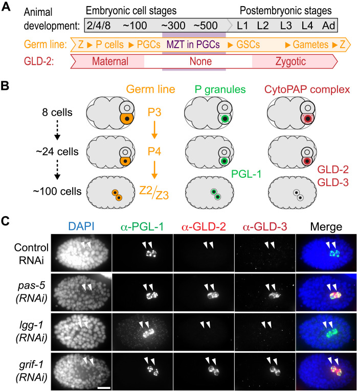
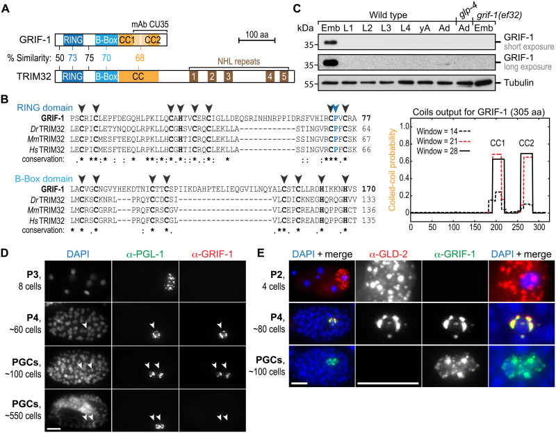
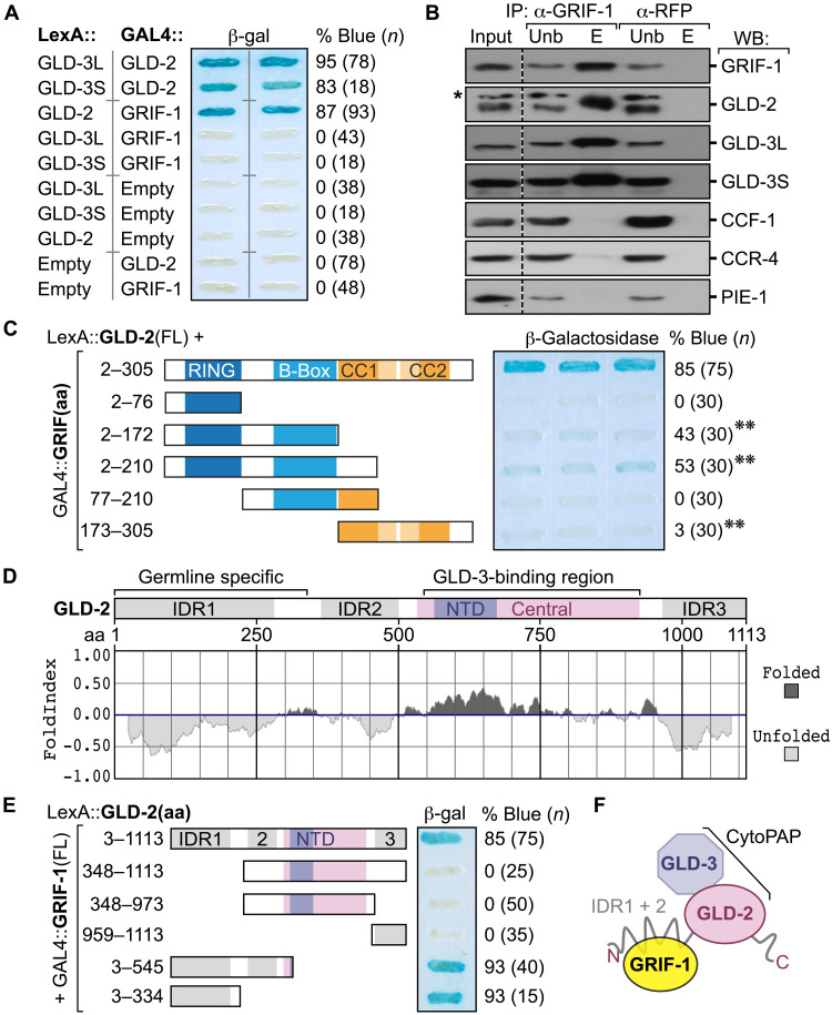
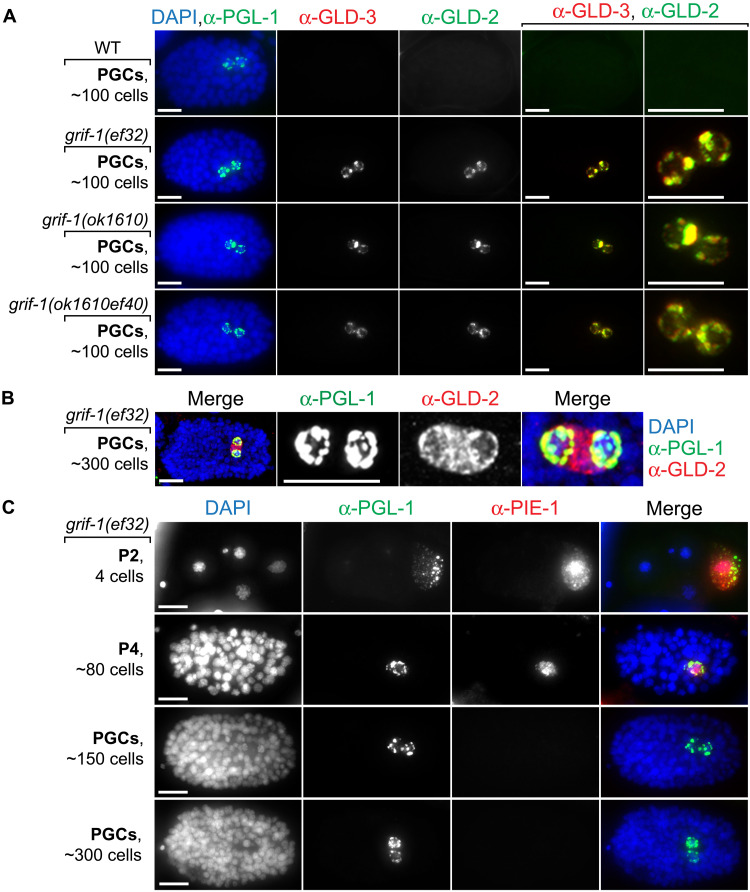
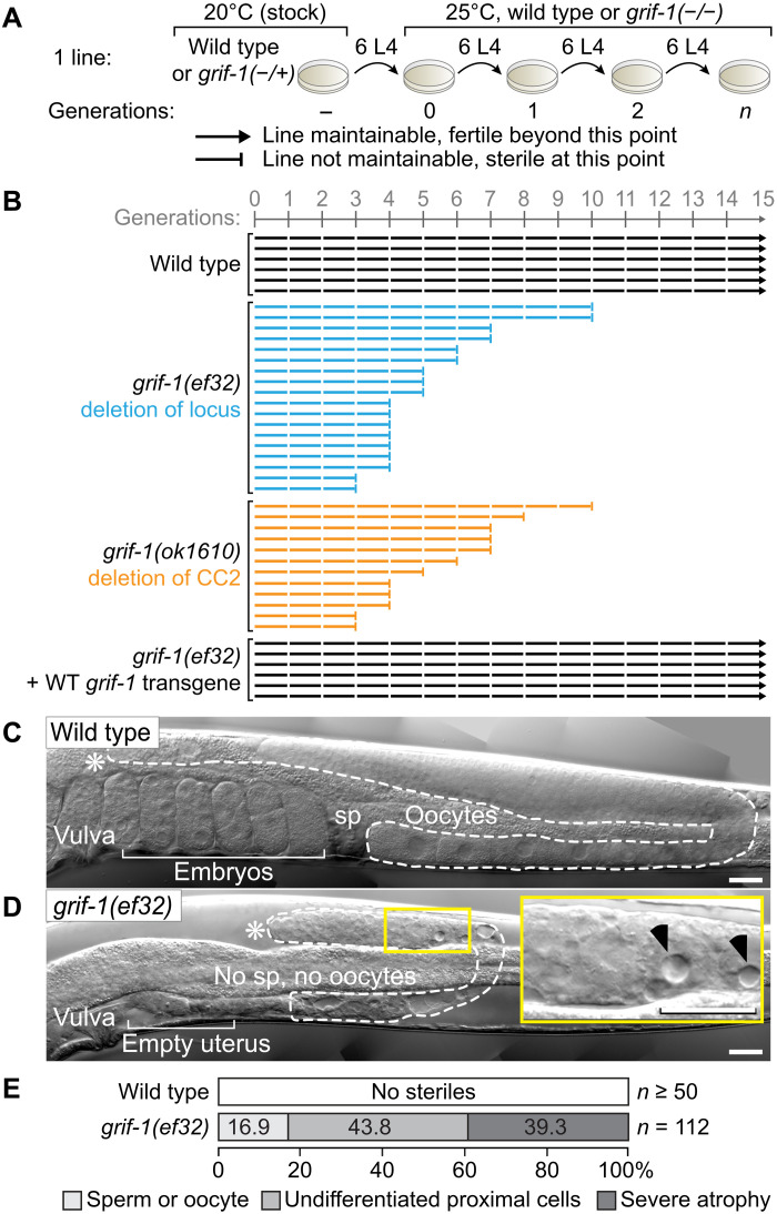
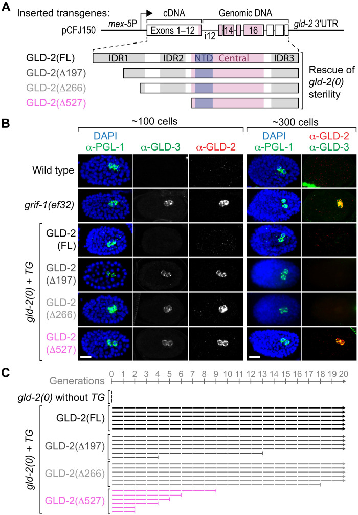
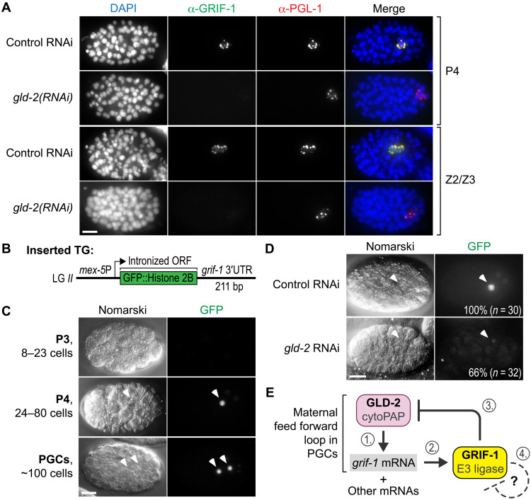
How the germ line achieves a clean transition from maternal to zygotic gene expression control is a fundamental problem in sexually reproducing organisms. Whereas several mechanisms terminate the maternal program in the soma, this combined molecular reset and handover are poorly understood for primordial germ cells (PGCs). Here, we show that GRIF-1, a TRIM32-related and presumed E3 ubiquitin ligase in Caenorhabditis elegans, eliminates the maternal cytoplasmic poly(A) polymerase (cytoPAP) complex by targeting the germline-specific intrinsically disordered region of its enzymatic subunit, GLD-2, for proteasome-mediated degradation. Interference with cytoPAP turnover in PGCs causes frequent transgenerational sterility and, eventually, germline mortality. Hence, positively acting maternal RNA regulators are cleared via the proteasome system to avoid likely interference between maternal and zygotic gene expression programs to maintain transgenerational fertility and acquire germline immortality. This strategy is likely used in all animals that preform their immortal germ line via maternally inherited germplasm determinants.


Figures

Fig. 1.
Fig. 1.. Maternal GLD-2 and GLD-3 are degraded in PGCs.
(A and B) Starting from fertilization (Z, zygote), maternal GLD-2 protein is specifically maintained in the germline (P) lineage, marked by the permanent P granule component PGL-1. During later embryonic stages, GLD-2 and its activator, GLD-3, are absent during the ZGA stages of either PGC (Z2 and Z3). Germline development continues postembryonically, and PGCs become dividing germline stem cells (GSCs). Zygotic GLD-2 abundance gradually increases in later larval (L) stages and remains high through late female gametogenesis. (C) Fixed ~100-cell stage wild-type embryos, treated with control RNAi, or targeting 26S proteasome (pas-5), autophagosome (lgg-1), or a RING finger gene (grif-1), stained for DNA [4,6-diamidino-2-phenylindole (DAPI)] and indicated proteins, whose given color code corresponds to the respective fluorescent detection channel (see Materials and Methods for further clarification). CytoPAP subunits GLD-2 and GLD-3 are stabilized in PGCs upon pas-5 (96%, n = 118) or grif-1 (100%, n > 100) but not control (100%, n > 90) or lgg-1 (100%, n > 100) knockdown. Merge, all channels. Scale bar, 10 μm.
Fig. 2.
Fig. 2.. GRIF-1 is a TRIM32-like protein abundant in PGCs.
(A and B) Across its primary sequence (top BLAST hit score of 1 × 10⁻⁶) and in its protein domain architecture, GRIF-1 is most similar to the E3 ubiquitin ligase TRIM32. However, its similarity is limited to the N-terminal half of TRIM32 because GRIF-1 lacks the additional RNA binding (NHL) domain. At its C terminus, GRIF-1 contains a noncontiguous CC domain (orange) predicted to be split into two parts (CC1 and CC2). (B) Multiple sequence alignments of the RING (blue) and single B-Box (cyan) domain. Despite several insertions, key amino acids [aa; cysteine (C) and histidine (H)] important for zinc finger formation (black arrowheads) or presumed E3 ligase activity [e.g., proline (P), blue arrowhead] are conserved: full stop (.), similar amino acid; colon (:), highly similar amino acid; asterisk (*), identical amino acid. Dark orange regions correspond to COILs (version 2.2) output, using strings of 14, 21, or 28 amino acids. (C to E) GRIF-1 expression is restricted to the embryonic germ line, temporarily overlapping with GLD-2 on P granules in P4 and early PGCs. (C) Western blots using mAb CU35 on extracts of wild-type larvae (L), adults without (yA) and with (Ad) embryos (Emb), and no germline (glp-4). (D and E) Fluorescent images of wild-type embryos (100%, n > 50 for each stage) stained with DAPI and antibodies against indicated proteins. Note that GRIF-1 expression always ceases in aged PGCs. Scale bars, 10 μm.
Fig. 3.
Fig. 3.. GRIF-1 interacts with the N-terminal IDR of GLD-2 cytoPAP.
(A) Beta-galactosidase (β-gal) activity (percentage of blue) of yeast colonies (n) coexpressing indicated fusion proteins; empty, no fusion. GRIF-1 interacts with GLD-2, but not with either isoform of GLD-3 (L or S), each binding to GLD-2. (B) Immunoprecipitated (IP) materials from embryonic extracts with antibodies directed against GRIF-1 or RFP that serves as a specificity control were probed in Western blots (WBs) for indicated proteins; 5% input (Inp) and unbound (Unb) materials and 50% of eluted (E) material loaded. Asterisk (*) shows unspecific background signal of anti–GLD-2 antibody. (C) Only FL GRIF-1 fused to GAL4 activation domain binds efficiently to GLD-2 in direct Y2H tests. Label as in (A); double asterisk (**) indicates colonies that became slightly blue after longer reaction times than in (A). (D) FoldIndex algorithm predicts three IDRs in GLD-2, including the entire N-terminal domain, encoded only in germline-specific transcripts of gld-2. The catalytical heart of GLD-2, the nucleotidyl-transferase domain (NTD), is embedded in a well-folded central domain that binds GLD-3 isoforms tightly for further enzymatic stimulation. (E) The germline-specific IDR of GLD-2 mediates the interaction with GRIF-1 in yeast. Label as in (A). (F) Working model of the interaction among GRIF-1 and GLD-2/3 cytoPAP subunits; GLD-3 represents either GLD-3L or GLD-3S.
Fig. 4.
Fig. 4.. Expression profile changes in grif-1 mutant PGCs.
(A to C) Fluorescent images of embryos at different developmental embryonic stages stained for DNA (DAPI) and indicated proteins. Merge, all channels. Scale bars, 10 μm. (A and B) GLD-2 and GLD-3 protein expression endures and remains P granular in freshly born PGCs of various grif-1 mutant ~100-stage embryos (100%, n > 100, each genotype). (B) GLD-2 redistributes from P granules in younger embryos to become strongly cytosolic in older grif-1 mutant embryos (100%, n > 100) correlating with minor genome activation at the ~300-cell stage in wild-type PGCs. (C) The maternal expression of the cytoplasmic RNA regulator and nucleoplasmic transcriptional repressor PIE-1 is, at large, unperturbed in grif-1(0) embryos and becomes undetectable beyond the 150-cell stage (see main text for details).
Fig. 5.
Fig. 5.. Transgenerational sterility and germline atrophy in grif-1 mutants.
(A) Experimental workflow to determine a mortal germline phenotype and legend to all data compilations as shown in (B). Six wild-type or grif-1(−/−) L4 animals, with or without expressing a wild-type (WT) grif-1 transgene, are passed across >15 generations at 25°C, starting from grif-1(+/−) heterozygotes, and maintained at 20°C. (B) A line (horizontal bar) is regarded as sterile (ending on a vertical bar) when mothers produce less than two progenies on average and further propagation became impossible. Wild-type or rescued grif-1 mutant animals continued to be fertile (ending on an arrowhead), and grif-1(−/−) mutant lines became completely sterile at early but varying generations. See fig. S6A for other grif-1 alleles and the rescuing transgene. (C and D) Nomarski images of a single gonadal arm (dotted line) in adults. Asterisks (*), distal end of gonad; self-fertile wild-type animals store sperm (sp) in their proximal spermatheca. Inset enlarges signatures of germline atrophy (black arrowheads); no differentiated gametes are present in this small gonad of a sterile grif-1 adult. Scale bars, 20 μm. (E) Sterile grif-1 animals display three major germ cell phenotypes in their gonads: defective gametes, no gametes but undifferentiated cells in the proximal region, and severe cell death and atrophy either proximally or throughout.
Fig. 6.
Fig. 6.. Prolonged GLD-2 expression in PGCs affects transgenerational fitness at 25°C.
(A) Stick diagrams of all transgene-expressed N-terminally truncated (Δ) GLD-2 protein variants used to rescue postembryonic germline development defects and early embryonic arrests of gld-2(0) mutant F1 animals and F2 embryos, respectively; labels as in Fig. 3D. Partially intronized (i) chromosome II–integrated gld-2 transgenes under the control of germ cell–specific mex-5 promoter (arrow) and cognate 3′ untranslated region (UTR) sequences. (B) Unlike in wild-type or FL GLD-2–rescued gld-2(0) animals and similar to PGCs in grif-1(ef32), PGCs of any GLD-2(ΔN terminus)–rescued gld-2(0) embryos display prolonged cytoPAP expression. Note that extended GLD-2 and GLD-3 perdurance is only maintained in PGCs of older grif-1(ef32) and GLD-2(Δ527aa) embryos (for quantitation, see main text). Scale bars, 10 μm. (C) Transgenerational defects are most severe upon removal of the entire IDR in GLD-2(Δ527aa). gld-2(0) transgenic animals analyzed and displayed as in Fig. 5. See also fig. S7 for extended analyses.
Fig. 7.
Fig. 7.. GRIF-1 expression depends on posttranscriptional positive regulation via GLD-2 cytoPAP.
(A) Fixed wild-type embryos of late P lineage stages, treated with low doses of RNAi targeting gld-2, and stained for DNA (DAPI) and indicated proteins. In rare escapers that also have mild gastrulation defects, endogenous GRIF-1 expression is compromised in PGL-1–positive P4 or PGC stages (100%, n = 20). Merge, all channels. Scale bar, 10 μm. (B) Cartoon display of strains harboring a genome-integrated green fluorescent protein (GFP) translational reporter transgene (TG) whose mRNA is transcribed from the germline-specific mex-5 promoter and posttranscriptionally regulated through the grif-1 3′UTR. The ORF of GFP is interrupted by three synthetic introns (not shown) and fused in frame to a histone 2B protein piece. Upon translation, the resulting fusion protein accumulates in the nucleus on chromatin. (C and D) Nomarski images of embryos at different developmental embryonic stages with corresponding autofluorescence of the translational reporter GFP signal (100%, n > 50). Scale bars, 10 μm. (C) Translational GFP reporter expression recapitulates developmental GRIF-1 expression profile. (D) RNAi knockdown compromises translational GFP reporter expression in many gld-2, but not in control, embryos. (E) Working model of GLD-2 cytoPAP–dependent grif-1 expression in P4 and early PGCs (steps 1 and 2). GRIF-1–mediated turnover of GLD-2 (step 3) stops this feed forward loop of maternal components also for other maternal—and potentially even zygotic—mRNA targets of GLD-2 cytoPAP. We hypothesize that a potential autoregulatory feedback loop of the presumed TRIM E3 ligase GRIF-1 may cause its elimination once its protein targets might be exhausted (step 4).

Similar articles

Cited by

References

    1. Strome S., Updike D., Specifying and protecting germ cell fate. Nat. Rev. Mol. Cell Biol. 16, 406–416 (2015). - PMC - PubMed
    1. Vastenhouw N. L., Cao W. X., Lipshitz H. D., The maternal-to-zygotic transition revisited. Development 146, dev161471 (2019). - PubMed
    1. Siddiqui N. U., Li X., Luo H., Karaiskakis A., Hou H., Kislinger T., Westwood J. T., Morris Q., Lipshitz H. D., Genome-wide analysis of the maternal-to-zygotic transition in Drosophila primordial germ cells. Genome Biol. 13, R11 (2012). - PMC - PubMed
    1. Voronina E., Seydoux G., Sassone-Corsi P., Nagamori I., RNA granules in germ cells. Cold Spring Harb. Perspect. Biol. 3, a002774 (2011). - PMC - PubMed
    1. Cao W. X., Kabelitz S., Gupta M., Yeung E., Lin S., Rammelt C., Ihling C., Pekovic F., Low T. C. H., Siddiqui N. U., Cheng M. H. K., Angers S., Smibert C. A., Wühr M., Wahle E., Lipshitz H. D., Precise temporal regulation of post-transcriptional repressors is required for an orderly drosophila maternal-to-zygotic transition. Cell Rep. 31, 107783 (2020). - PMC - PubMed

MeSH terms