. 2022 Oct 18;119(42):e2123338119.
doi: 10.1073/pnas.2123338119. Epub 2022 Oct 14.

5-methylcytosine (m5C) RNA modification controls the innate immune response to virus infection by regulating type I interferons

Affiliations

5-methylcytosine (m5C) RNA modification controls the innate immune response to virus infection by regulating type I interferons

Yuexiu Zhang et al. Proc Natl Acad Sci U S A. .

Abstract

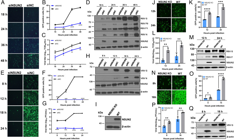
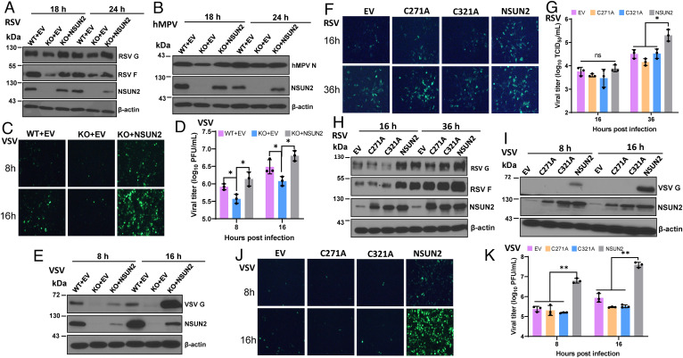
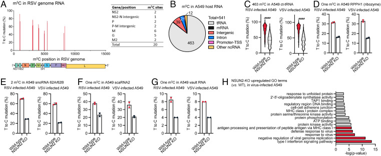
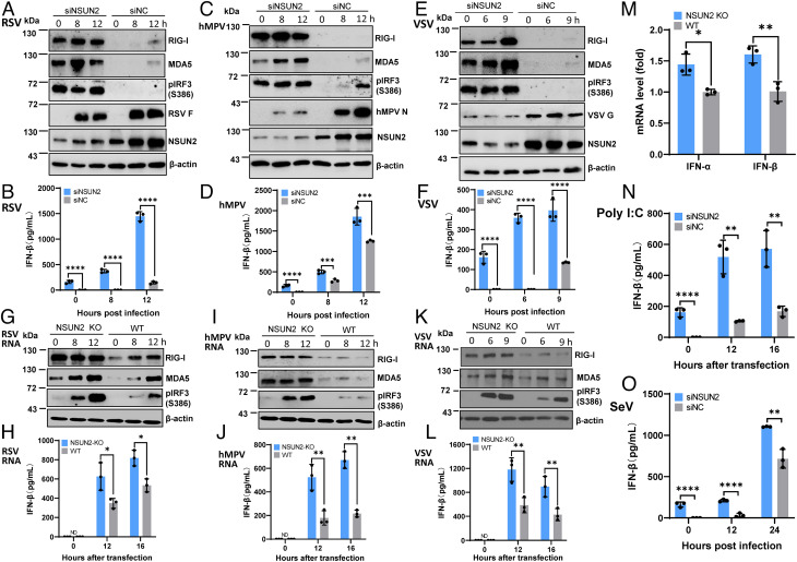
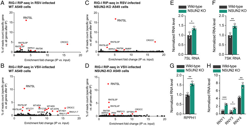
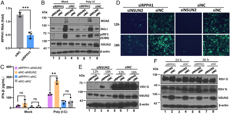
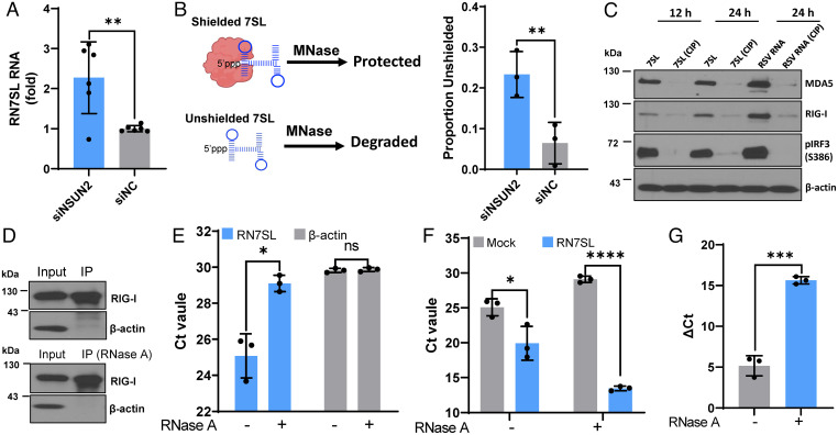
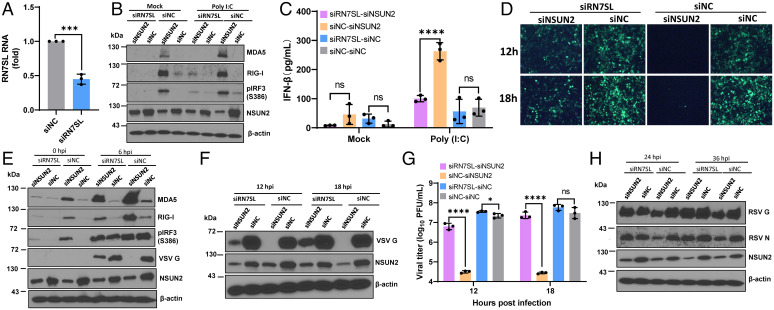
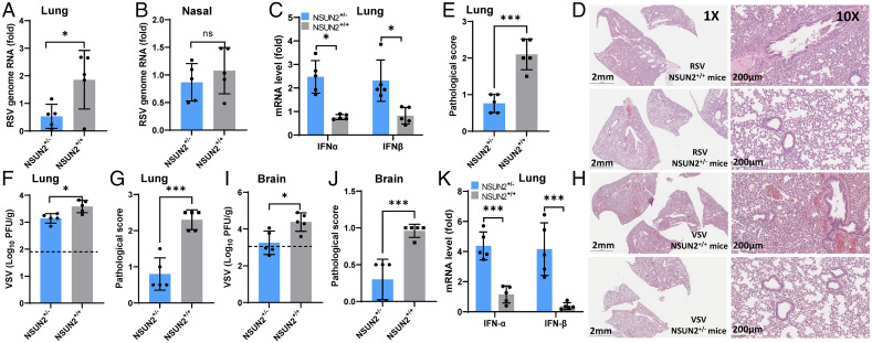
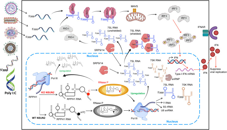
5-methylcytosine (m5C) is one of the most prevalent modifications of RNA, playing important roles in RNA metabolism, nuclear export, and translation. However, the potential role of RNA m5C methylation in innate immunity remains elusive. Here, we show that depletion of NSUN2, an m5C methyltransferase, significantly inhibits the replication and gene expression of a wide range of RNA and DNA viruses. Notably, we found that this antiviral effect is largely driven by an enhanced type I interferon (IFN) response. The antiviral signaling pathway is dependent on the cytosolic RNA sensor RIG-I but not MDA5. Transcriptome-wide mapping of m5C following NSUN2 depletion in human A549 cells revealed a marked reduction in the m5C methylation of several abundant noncoding RNAs (ncRNAs). However, m5C methylation of viral RNA was not noticeably altered by NSUN2 depletion. In NSUN2-depleted cells, the host RNA polymerase (Pol) III transcribed ncRNAs, in particular RPPH1 and 7SL RNAs, were substantially up-regulated, leading to an increase of unshielded 7SL RNA in cytoplasm, which served as a direct ligand for the RIG-I-mediated IFN response. In NSUN2-depleted cells, inhibition of Pol III transcription or silencing of RPPH1 and 7SL RNA dampened IFN signaling, partially rescuing viral replication and gene expression. Finally, depletion of NSUN2 in an ex vivo human lung model and a mouse model inhibits viral replication and reduces pathogenesis, which is accompanied by enhanced type I IFN responses. Collectively, our data demonstrate that RNA m5C methylation controls antiviral innate immunity through modulating the m5C methylome of ncRNAs and their expression.

Keywords: 5-methylcytosine; innate immune response; interferon; virus infection.


Conflict of interest statement

Competing interest statement: C.H. is a scientific founder of Accent Therapeutics, Ins. J.L. and C.H. are filing a provision patent application, 21-T-003/IDF050556.

Figures

Fig. 1.
Fig. 1.
NSUN2 depletion suppresses viral replication and gene expression. NSUN2 depletion suppresses RSV (AD) and VSV (EH) replication. A549 cells were transfected with siNSUN2 or siRNA negative control (siNC). After 24 h, cells were infected with GFP-expressing viruses (rgRSV or rVSV-GFP) at an MOI of 0.1, GFP expression was captured by fluorescence microscopy (A and E), and GFP-positive cells were quantified by flow cytometry (B and F). A single-step growth curve shows the release of RSV (C) and VSV (G). Total cell extracts were harvested from rgRSV (D)- or rVSV-GFP (H)–infected A549 cells and subjected to Western blot (D and H). (I) Western blot showing KO of NSUN2 in A549 cells. (JQ) NSUN2 KO suppresses RSV and VSV replication. NSUN2-KO A549 cells or control sgRNA A549 cells were infected with rgRSV (JM) or rVSV-GFP (NQ) at an MOI of 0.1. GFP images were captured (J and N), percent of GFP-positive cells was quantified (K and O), RSV (M) or VSV (Q) protein expression was determined by Western blot, and RSV (L) and VSV (P) titers were determined. All results are from three independent experiments. Data were analyzed using Student’s t test (*P < 0.05, **P < 0.01, ***P < 0.001, ****P < 0.0001).
Fig. 2.
Fig. 2.
m5C methyltransferase of NSUN2 is essential for its proviral activity. (AE) Rescue of viral replication and gene expression by transfection of NSUN2 plasmid. NSUN2-KO or control A549 cells were transfected with 1 μg of empty vector (EV) pCAGGS or pCAGGS-NSUN2, followed by infection with rgRSV (A), hMPV (B), or VSV (CE) at an MOI of 1.0. Transfection of pCAGGS-NSUN2 rescued RSV (A), hMPV (B), and VSV (E) protein expression, as well as GFP expression by rVSV-GFP (C) and VSV titer (D). (FK) Two cysteine residues (C271 and C321) in NSUN2 protein are essential for its proviral activity. NSUN2-KO or control A549 cells were transfected with pCAGGS-NSUN2 or mutants followed by infection with rgRSV (F, G, and H) or rVSV-GFP (I, J, and K) at an MOI of 1.0. Transfection of WT NSUN2 but not NSUN2-C271A or NSUN2-C321A rescued the GFP expression (F and J), viral titer (G and K), and viral protein expression (H and I) in NSUN2-KO cells. All results are from three independent experiments. Data were analyzed using s Student’s t test (*P < 0.05; **P < 0.01).
Fig. 3.
Fig. 3.
NSUN2 depletion leads to an enhanced up-regulation of type I IFN signaling and antivirus immune response, in which it also controls the m5C methylation in some A549 host RNAs. (A) (Left) location of 20 m5C sites within RSV genome RNA. The x axis shows the m5C site position in RSV genome; the y axis shows the m5C site mutation level. (Right) Number of m5C sites in each RSV gene. (B) Distribution of NSUN2-regulated m5C sites in major RNA species from A549 host RNA (shared by both RSV-infected and VSV-infected cells). (C) Mutation levels of NSUN2-regulated m5C sites in cytoplasmic tRNAs from NSUN2-KO A549 cells vs. WT cells. P values were determined using two-tailed t test for paired samples (****P < 0.0001). (D–G) Mutation levels of one m5C site in RPPH1 (D), two m5C sites in snoRNA 62A and 62B (E), one m5C site in scaRNA2 (F), and one m5C site in vault RNA (vtRNA1-1) (G) from NSUN2-KO A549 cells vs. WT cells. For (D)–(G), mean values ± SD are shown; n = 2. (H) Enriched GO clusters of up-regulated genes in virus-infected NSUN2-KO A549 cells (vs. virus-infected WT A549 cells). The clusters are ranked by P value.
Fig. 4.
Fig. 4.
NSUN2 depletion leads to the activation of a higher type I IFN signaling pathway. (AF) NSUN2 depletion induces a higher type I IFN after virus infection. A549 cells were transfected with siNSUN2 or siNC, and were infected with rgRSV (A and B), hMPV (C and D), or rVSV-GFP (E and F) at an MOI of 1.0, 5.0, and 1.0, respectively. Cell lysates were subjected to Western blot analyses (A, C, and E). IFN-β in cell culture supernatants was detected by ELISA (B, D, and F). (GL) NSUN2 KO induces a higher type I IFN after transfection with viral RNA. NSUN2-KO or control A549 cells were transfected with virion RNAs (2 × 10⁶ copies/well) of rgRSV (G and H), hMPV (I and J), or rVSV-GFP (K and L). ND indicates the value was below the detection limit (50 pg/mL). (M) Quantification of IFN mRNA by RT-qPCR. Total RNA was extracted from NSUN2-KO or control sgRNA-treated A549 cells. IFN-α and IFN-β mRNA was quantified by RT-qPCR. β-actin was used for internal control. (N) NSUN2 KO induces a higher type I IFN after transfection with poly(I:C). Cells were transfected with 0.5 µg/well of poly(I:C). (O) NSUN2 KO induces a higher type I IFN after infection with rSeV-GFP. An MOI of 1.0 was used for infection. Data were analyzed using a Student’s t test (*P < 0.05, **P < 0.01, ***P < 0.001, ****P < 0.0001).
Fig. 5.
Fig. 5.
m5C deficiency-mediated antiviral response is dependent on RIG-I but not MDA5. Confluent WT A549 cells and RIG-I-KO (RIG-I−/−) A549 cells were transfected with siRNA against NSUN2 or control siRNA, followed by infection with rgRSV at an MOI of 0.1. GFP images were taken by fluorescence microscopy (A) and viral titer in cell culture supernatants was determined by a 50% tissue culture infective dose (TCID50) (B). (C) A549 cells were transfected with siRNA against NSUN2, MDA5, or both, followed by infection with rgRSV at an MOI of 0.1, and viral titer in cell culture supernatants was determined by TCID50 . Data were analyzed using a Student’s t test (*P < 0.05, **P < 0.01, ***P < 0.001).
Fig. 6.
Fig. 6.
RIG-I binds to 7SL RNA (RN7SL) in vivo when overexpressing RIG-I in A549 cells, and NSUN2 depletion leads to elevated Pol III transcription of 7SL, 7SK, RPPH1, and Y RNA. Enriched genes in RIG-I RIP-seq when overexpressing RIG-I in RSV (A)- or VSV (B)-infected WT A549 cells. Enriched genes in RIG-I RIP-seq when overexpressing RIG-I in RSV (C)- or VSV (D)-infected NSUN2-KO A549 cells. For (A)–(D), the y axis shows the percentage of how much the reads count from one specific gene occupies in all IP-enriched genes; the x axis shows the enrichment fold change after IP (vs. input). (E–H) Relative RNA levels of 7SL (E), 7SK (F), RPPH1 (G), and Y (H) RNAs in NSUN2-KO A549 cells vs. the WT A549 cells without viral infection, which are transcribed by Pol III. For (E)(H), mean values ± SD are shown; n = 3 biologically independent samples. P values were determined using an unpaired t test (*P < 0.05, **P < 0.01, ***P < 0.001).
Fig. 7.
Fig. 7.
Silencing of RPPH1 rescues virus replication. (A) Knockdown of RPPH1 by siRNA. A549 cells were transfected with control siRNA or siRNA against RPPH1, and RPPH1 RNA was quantified by RT-qPCR. (B and C) Silencing of RPPH1 attenuates IFN signaling. A549 cell were transfected with siNSUN2/siRPPH1, siNSUN2/siNC, siRPPH1/siNC, or siNC, followed by transfection with poly(I:C), and cell lysate and supernatant were harvested for Western blot (B) and ELISA (C) at 0 and 8 h after transfection. (DF) Silencing of RPPH1 partially rescues VSV and RSV replication. A549 cells were treated with siRNAs described in (B), followed by infection with rVSV-GFP (D and E) and rgRSV (F) at an MOI of 0.1; representative GFP images in rVSV-GFP–infected cells were monitored (D). Cell lysates were prepared for Western blot against VSV (E) or RSV (F) proteins. Data were analyzed using Student’s t test (**P < 0.01, ***P < 0.001).
Fig. 8.
Fig. 8.
7SL RNAs are the direct ligands of RIG-I. (A) Total 7SL RNAs are elevated in NSUN2 knockdown cells. A549 cells were transfected with siNSUN2 or siNC. At 24 h posttransfection, 7SL RNA was quantified by RT-qPCR and normalized by β-actin mRNA. Data shown are fold increases in NSUN2-depleted cells relative to the control siRNA-treated cells. (B) Unshielded 7SL RNAs are elevated in NSUN2 knockdown cells. A549 cells were transfected with siNSUN2 or siNC, incubated with or without MNase, and the unshielded 7SL RNAs were measured by RT-qPCR. The 7SL RNAs were normalized by β-actin mRNA. Data shown are unshielded 7SL RNA relative to total 7SL RNA. (C) 7SL RNAs activate RIG-I signaling. A549 cells were transfected with in vitro–transcribed 7SL RNA or RSV virion RNA pretreated with or without CIP. The cell lysates were harvested for Western blot. (DG) Pulldown of 7SL RNA by RIG-I. HEK293T cells were transfected with FLAG–RIG-I plasmid, and the input and immunoprecipitated RIG-I proteins were analyzed by Western blot (D). The purified FLAG–RIG-I proteins were treated with or without RNase A to remove endogenous 7SL RNA (E). The purified FLAG–RIG-I proteins with or without RNase A treatment were inoculated with or without in vitro–transcribed 7SL RNA, and the level of 7SL RNA bound to RIG-I was measured by RT-qPCR (F and G). Data are presented as cycle threshold (Ct) (F) and ΔCt (ΔCt = CtMock − Ct7SL) (G). All data are from three (n = 3) independent experiments (except A, n = 6). Data were analyzed using a Student’s t test (*P < 0.05, **P < 0.01, ***P < 0.001, ****P < 0.0001).
Fig. 9.
Fig. 9.
Silencing of 7SL RNA (RN7SL) rescues virus replication. (A) Efficient knockdown of 7SL RNA using siRNA. A549 cells were transfected with siRNA against 7SL or control siRNA. At 24 h later, 7SL RNA was quantified by RT-qPCR. (B and C) Silencing of 7SL RNA attenuates IFN signaling upon poly(I:C) stimulation. A549 cells were transfected with siNSUN2/si7SL, siNSUN2/siNC, si7SL/siNC, or siNC, followed by transfection with poly(I:C), and cell lysates were harvested for Western blot (B) and supernatants were collected for measurement of IFN-β by ELISA (C) at 0 and 8 h after transfection. (DG) Silencing of 7SL RNA partially rescues VSV replication. A549 cells were transfected with siNSUN2/si7SL, siNSUN2/siNC, si7SL/siNC, or siNC, followed by infection with rVSV-GFP at an MOI of 0.1. GFP images were taken by fluorescence microscopy (D). At early (0 and 6 h) (E) and late (12 and 18 h) (F) time points after infection, cell lysates were harvested for Western blot. VSV titer was rescued in NSUN2 and 7SL double-silenced A549 cells (G). (H) Silencing of 7SL RNA partially rescues RSV replication. A549 cells were treated with siRNA described in (D) and infected with rgRSV at an MOI of 0.1; cell lysates were subjected to Western blot. All data are from three (n = 3) independent experiments. Data were analyzed using a Student’s t test (*P < 0.05, ***P < 0.001, ****P < 0.0001).
Fig. 10.
Fig. 10.
Knockout of NSUN2 in mice reduces viral replication and enhances type I IFN responses. (AE) RSV infection in NSUN2+/- and WT (NSUN2+/+) mice. The 4-6-wk-old specific pathogen-free (SPF) female WT mice and NSUN2+/- mice were intranasally inoculated with 4 × 10⁵ TCID50 of rgRSV. At day 3 postinoculation, mice were euthanized and total RNA was extracted from the right lung (A) and the nasal turbinate (B) from each mouse for quantification of RSV genome RNA and type I IFN (IFN-α and IFN-β) mRNA in lungs (C) by real-time RT-PCR. Representative lung histological images (D) and average lung histology score from each group are shown (E). Each slide was scored based on severity of lesion. Scores were as follows: 0, no lesion; 1, mild; 2, moderate; 3, severe. (FK) VSV infection in NSUN2+/- and WT mice. The 4-6-wk-old SPF female WT mice and NSUN2+/- mice were intranasally inoculated with 106 plaque-forming units (PFU) of VSV. At day 3 postinoculation, all mice were euthanized, and VSV titers in lungs (F) and brain (I) were quantified by plaque assay. Total RNA was extracted from lung tissues for quantification of IFN-α and IFN-β mRNA by RT-qPCR (K). Representative lung histological images (H) and average score (G) from each group are shown. Average brain histology score from each group is shown (J). Scores were based on the severity of encephalitis. Scores were as follows: 0, no lesion; 1, mild; 2, moderate; 3, severe. Data were analyzed using a Student’s t test (*P < 0.05, ***P < 0.001).
Fig. 11.
Fig. 11.
Model of NSUN2 regulation of type I IFN signaling. Upon depletion of NSUN2 in A549 cells, the increase in RPPH1 expression and the decrease in m5C modification in RPPH1 may lead to enhance Pol III activity via regulating RNase P enzymatic activity. The elevation of Pol III–transcribed RNAs, especially 7SL RNAs, leads to an increase in unshielded 7SL RNAs in the cytoplasm, which can be recognized by RIG-I, causing type I IFN signaling. Upon virus infection, the increased PRRs quickly recognize the invading non-self RNAs, further amplifying IFN signaling and leading to a dramatically enhanced type I IFN which in turn inhibits virus propagation.

References

    1. Li Q., et al. , NSUN2-mediated m5C methylation and METTL3/METTL14-mediated m6A methylation cooperatively enhance p21 translation. J. Cell. Biochem. 118, 2587–2598 (2017). - PMC - PubMed
    1. Zhao B. S., Roundtree I. A., He C., Post-transcriptional gene regulation by mRNA modifications. Nat. Rev. Mol. Cell Biol. 18, 31–42 (2017). - PMC - PubMed
    1. Fu Y., Dominissini D., Rechavi G., He C., Gene expression regulation mediated through reversible m6A RNA methylation. Nat. Rev. Genet. 15, 293–306 (2014). - PubMed
    1. Lu M., et al. , N6-methyladenosine modification enables viral RNA to escape recognition by RNA sensor RIG-I. Nat. Microbiol. 5, 584–598 (2020). - PMC - PubMed
    1. Baer M., Nilsen T. W., Costigan C., Altman S., Structure and transcription of a human gene for H1 RNA, the RNA component of human RNase P. Nucleic Acids Res. 18, 97–103 (1990). - PMC - PubMed

Publication types

LinkOut - more resources