. 2023 Jan 10;7(1):167-173.
doi: 10.1182/bloodadvances.2022008141.

Integrated flow cytometry and sequencing to reconstruct evolutionary patterns from dysplasia to acute myeloid leukemia

Affiliations

Integrated flow cytometry and sequencing to reconstruct evolutionary patterns from dysplasia to acute myeloid leukemia

Catia Simoes et al. Blood Adv. .

Abstract

Clonal evolution in acute myeloid leukemia (AML) originates long before diagnosis and is a dynamic process that may affect survival. However, it remains uninvestigated during routine diagnostic workups. We hypothesized that the mutational status of bone marrow dysplastic cells and leukemic blasts, analyzed at the onset of AML using integrated multidimensional flow cytometry (MFC) immunophenotyping and fluorescence-activated cell sorting (FACS) with next-generation sequencing (NGS), could reconstruct leukemogenesis. Dysplastic cells were detected by MFC in 285 of 348 (82%) newly diagnosed patients with AML. Presence of dysplasia according to MFC and World Health Organization criteria had no prognostic value in older adults. NGS of dysplastic cells and blasts isolated at diagnosis identified 3 evolutionary patterns: stable (n = 12 of 21), branching (n = 4 of 21), and clonal evolution (n = 5 of 21). In patients achieving complete response (CR), integrated MFC and FACS with NGS showed persistent measurable residual disease (MRD) in phenotypically normal cell types, as well as the acquisition of genetic traits associated with treatment resistance. Furthermore, whole-exome sequencing of dysplastic and leukemic cells at diagnosis and of MRD uncovered different clonal involvement in dysplastic myelo-erythropoiesis, leukemic transformation, and chemoresistance. Altogether, we showed that it is possible to reconstruct leukemogenesis in ∼80% of patients with newly diagnosed AML, using techniques other than single-cell multiomics.


Conflict of interest statement

Conflict-of-interest disclosure: F.P. received honoraria and research funding from Oryzon, Janssen, Bristol-Myers Squibb (BMS)/Celgene. R.A. serves as a member of the board of directors advisory committees for Incyte Corporation and Astellas and received honoraria from Novartis, Celgene, and Incyte. J.A.P.-S. received honoraria and funding budget for research projects and is active on the advisory board and learning activities or conferences of Janssen, Takeda, Pfizer, Jazz, BMS, Amgen, and Gilead. J.F.S.-M. does consultancy and is a member of the board of directors advisory committees for AbbVie, Amgen, BMS, Celgene, GlaxoSmithKline (GSK), Janssen, Karyopharm, Merck Sharpe & Dohme, Novartis, Regeneron, Roche, Sanofi, SecuraBio, and Takeda. P.M. provides consultancy, is a member of the board of directors advisory committees and speaker’s bureau, and received research funding from Celgene, Sanofi, Incyte, Karyopharm, Novartis, Stemline/Menarini, Agios, Astellas Pharma, and Daiichi Sankyo; is also a member of the board of directors advisory committees for Pfizer, Teva, and AbbVie; received research funding from and is a member of speaker’s bureau for Janssen; and provides consultancy for Tolero Pharmaceutical, Forma Therapeutics, and Glycomimetics. B.P. served as a consultant and received honoraria from Adaptive, Amgen, BD Biosciences, BMS/Celgene, GSK, Janssen, Roche, Sanofi, and Takeda and received research support from BMS/Celgene, GSK, Roche, Sanofi, and Takeda. The remaining authors declare no competing financial interests.

Figures

None
Graphical abstract
Figure 1.
Figure 1.
Reconstructing clonal evolution from dysplasia to AML. (A) Study design. BM aspirates collected from 358 patients with newly diagnosed AML were analyzed using MFC. In 21 patients, leukemic cells were isolated using FACS according to patient-specific aberrant phenotypes and so were cells of the neutrophil (Neutro), monocytic (Mono), and erythroid (Erythro) lineages whenever dysplastic phenotypes were observed in 1 or more lineages. T cells were systematically isolated for germline DNA. NGS of genes frequently mutated in myeloid neoplasms was performed in all cell types available in each patient. (B) Mutational status of 33 genes in T cells (T), neutrophils (N), monocytes (M), erythroblasts (E), and blasts (B) isolated from 21 patients at the onset of AML; cell types not available for NGS are represented with gray lines. Mutated genes were colored in a gradient of red according to the variant allele frequency (VAF). Three models were identified: (1) stable transition according to identical mutational landscapes in blasts and dysplastic cells; (2) branching evolution with blasts originating from leukemic stem cells other than the ones driving dysplasia, due to mutations absent in blasts and present in dysplastic cells; and (3) clonal evolution with new mutations in blasts onto mutations shared between these and dysplastic cells. (C) Percentage of mutations grouped according to functional categories that were simultaneously mutated in mature dysplastic cells and blasts (gray), or that were private in the former (blue) and the latter (purple). Functional categories with significantly different distributions were highlighted in bold.
Figure 1.
Figure 1.
Reconstructing clonal evolution from dysplasia to AML. (A) Study design. BM aspirates collected from 358 patients with newly diagnosed AML were analyzed using MFC. In 21 patients, leukemic cells were isolated using FACS according to patient-specific aberrant phenotypes and so were cells of the neutrophil (Neutro), monocytic (Mono), and erythroid (Erythro) lineages whenever dysplastic phenotypes were observed in 1 or more lineages. T cells were systematically isolated for germline DNA. NGS of genes frequently mutated in myeloid neoplasms was performed in all cell types available in each patient. (B) Mutational status of 33 genes in T cells (T), neutrophils (N), monocytes (M), erythroblasts (E), and blasts (B) isolated from 21 patients at the onset of AML; cell types not available for NGS are represented with gray lines. Mutated genes were colored in a gradient of red according to the variant allele frequency (VAF). Three models were identified: (1) stable transition according to identical mutational landscapes in blasts and dysplastic cells; (2) branching evolution with blasts originating from leukemic stem cells other than the ones driving dysplasia, due to mutations absent in blasts and present in dysplastic cells; and (3) clonal evolution with new mutations in blasts onto mutations shared between these and dysplastic cells. (C) Percentage of mutations grouped according to functional categories that were simultaneously mutated in mature dysplastic cells and blasts (gray), or that were private in the former (blue) and the latter (purple). Functional categories with significantly different distributions were highlighted in bold.
Figure 2.
Figure 2.
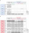
Different clonal involvement in dysplastic myelo-erythropoiesis, leukemic transformation, and chemoresistance. (A-B) Patients (Pat.) with undetectable (n = 5) and persistent (n = 5) MRD had phenotypically normal CD34+ HPCs and MRD cells, respectively isolated, whereas cells of the neutrophil, monocyte, and erythroid lineages were isolated in all patients. NGS of genes frequently mutated in myeloid neoplasms was performed in all cell types available in each patient. The VAF of mutated genes was colored in a gradient of gray. If 2 mutations in the same gene were detected, the 2 VAFs are indicated. (C) Representative patient with exome sequencing of dysplastic cell types, blasts at diagnosis, and persistent MRD. The fish plot illustrates different clonal compositions at different stages of disease progression. The blue arrow is pointing to a mutation present in dysplastic cells though absent in blasts at diagnosis and MRD, the olive arrow is pointing to mutations present in dysplastic cells and blasts at diagnosis though not at MRD, and the black arrow points to mutations present in MRD and dysplastic cells though not in blasts at diagnosis. The bar widths indicate the respective VAFs.
Figure 2.
Figure 2.
Different clonal involvement in dysplastic myelo-erythropoiesis, leukemic transformation, and chemoresistance. (A-B) Patients (Pat.) with undetectable (n = 5) and persistent (n = 5) MRD had phenotypically normal CD34+ HPCs and MRD cells, respectively isolated, whereas cells of the neutrophil, monocyte, and erythroid lineages were isolated in all patients. NGS of genes frequently mutated in myeloid neoplasms was performed in all cell types available in each patient. The VAF of mutated genes was colored in a gradient of gray. If 2 mutations in the same gene were detected, the 2 VAFs are indicated. (C) Representative patient with exome sequencing of dysplastic cell types, blasts at diagnosis, and persistent MRD. The fish plot illustrates different clonal compositions at different stages of disease progression. The blue arrow is pointing to a mutation present in dysplastic cells though absent in blasts at diagnosis and MRD, the olive arrow is pointing to mutations present in dysplastic cells and blasts at diagnosis though not at MRD, and the black arrow points to mutations present in MRD and dysplastic cells though not in blasts at diagnosis. The bar widths indicate the respective VAFs.

References

    1. Benard BA, Leak LB, Azizi A, Thomas D, Gentles AJ, Majeti R. Clonal architecture predicts clinical outcomes and drug sensitivity in acute myeloid leukemia. Nat Commun. 2021;12(1):1–13. - PMC - PubMed
    1. Miles LA, Bowman RL, Merlinsky TR, et al. Single-cell mutation analysis of clonal evolution in myeloid malignancies. Nature. 2020;587(7834):477–482. - PMC - PubMed
    1. Morita K, Wang F, Jahn K, et al. Clonal evolution of acute myeloid leukemia revealed by high-throughput single-cell genomics. Nat Commun. 2020;11(1):1–17. - PMC - PubMed
    1. Lindsley RC, Mar BG, Mazzola E, et al. Acute myeloid leukemia ontogeny is defined by distinct somatic mutations. Blood. 2015;125(9):1367–1376. - PMC - PubMed
    1. Weinberg OK, Pozdnyakova O, Campigotto F, et al. Reproducibility and prognostic significance of morphologic dysplasia in de novo acute myeloid leukemia. Mod Pathol. 2015;28(7):965–976. - PubMed

Publication types