Review
. 2022 Oct 13;185(21):3857-3876.
doi: 10.1016/j.cell.2022.09.023.

The JAK-STAT pathway at 30: Much learned, much more to do

Affiliations
Review

The JAK-STAT pathway at 30: Much learned, much more to do

Rachael L Philips et al. Cell. .

Abstract

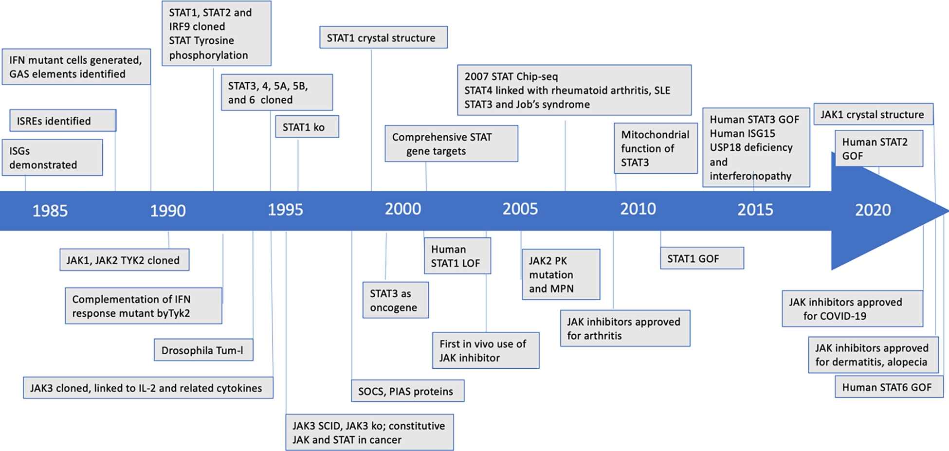
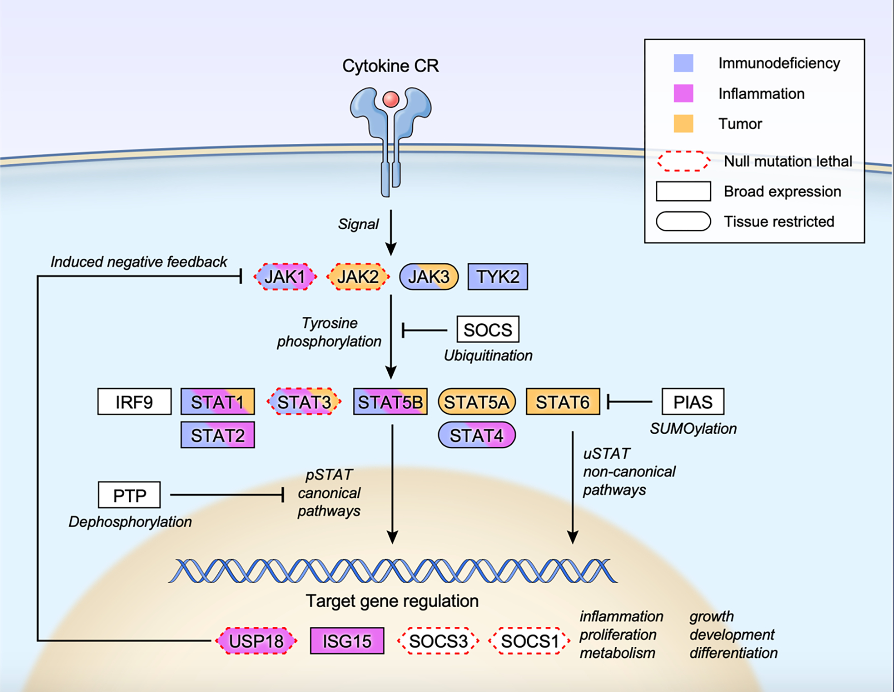
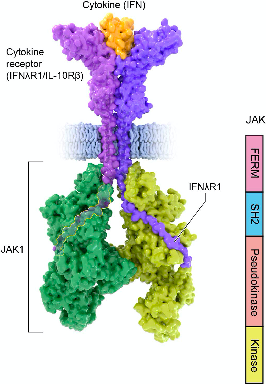
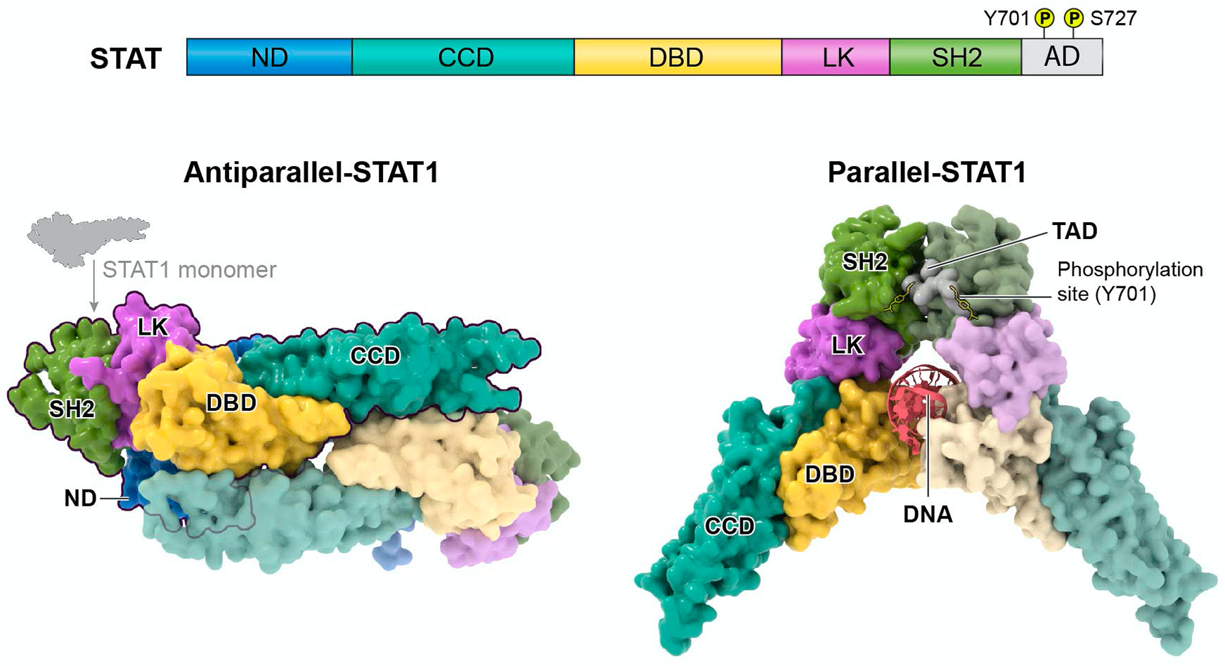
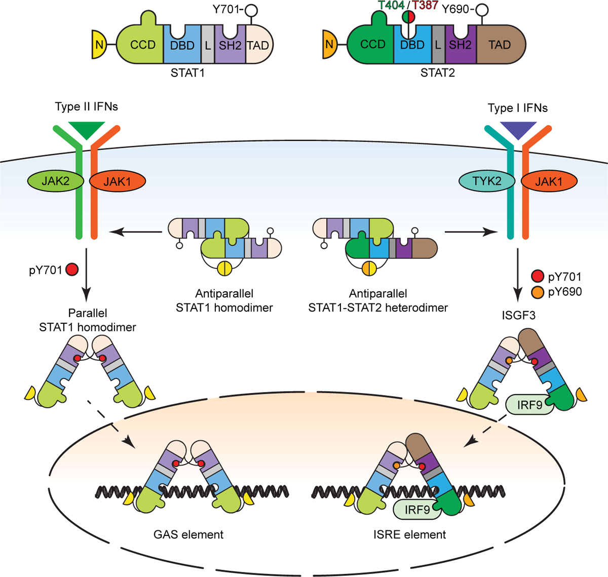
The discovery of the Janus kinase (JAK)-signal transducer and activator of transcription (STAT) pathway arose from investigations of how cells respond to interferons (IFNs), revealing a paradigm in cell signaling conserved from slime molds to mammals. These discoveries revealed mechanisms underlying rapid gene expression mediated by a wide variety of extracellular polypeptides including cytokines, interleukins, and related factors. This knowledge has provided numerous insights into human disease, from immune deficiencies to cancer, and was rapidly translated to new drugs for autoimmune, allergic, and infectious diseases, including COVID-19. Despite these advances, major challenges and opportunities remain.


Conflict of interest statement

Declaration of interests The NIH holds a US patent related to JAK inhibitors, and Dr. O’Shea receives royalty income.

Figures

Figure 1.
Figure 1.. JAK-STAT Timeline.
This timeline highlights various milestones in understanding the JAK-STAT pathway since 1984. GAS, gamma activated sequence; IFN, interferon; ISGs, IFN stimulated genes, ISRE, interferon-sensitive response element; SCID, severe combined immune deficiency; KO, knockout; SLE, systemic lupus erythematosus; PK, pseudokinase; MPN, myeloproliferative neoplasms; GOF, gain-of-function; SOCS, suppressor of cytokine signaling; PIAS, protein inhibitor of activated STAT, Tum-l, tumorous lethal.
Figure 2.
Figure 2.. Overall scheme of JAK STAT signaling.
Cytokine-receptor engagement transmits signal to tyrosine phosphorylate and activate receptor associated JAKs, which subsequently phosphorylate and activate STATs. Tyrosine phosphorylated STATs (pSTATs) form dimers, translocate to the nucleus, bind to target DNA sequences, and regulate gene expression (pSTAT canonical pathways). Unphosphorylated STATs also form dimers, enter the nucleus and regulate transcription (uSTAT non-canonical pathways). Multiple inducible negative feedback systems are in place to constrain the signaling circuit that include SOCS (suppressor of cytokine signaling) family proteins, PIAS (protein inhibitor of activated STAT) family proteins, PTP (protein tyrosine phosphatase), USP18 and ISG15. Human monogenic mutations that lead to gain-of-function and/or loss-of-function phenotypes are color coded as immunodeficiency (blue), inflammation (pink) or tumor (orange) for each constituent gene. Genes whose null mutation in mice leads to lethality is marked with red dotted border (Mouse Genome Informatics; http://www.informatics.jax.org). Tissue expression pattern of each protein is also marked as broad expression (square border) or tissue restricted (oval border) (The human protein atlas; https://www.proteinatlas.org/search).
Figure 3.
Figure 3.. Structure of Janus kinases.
Structure of activated Janus Kinase dimer (green; PDB 7T6F) complexed with the intracellular domain of matured IFNlR1/IL-10Rb (purple) bound to IFN-l (orange) (PDB 5T5W). Of note, the intracellular portion of the receptor binds JAK1 FERM and SH2 domains through N-terminal Box1 and C-terminal Box2 motifs. The JAK kinase-like or pseudokinase domain promotes dimerization of the cytokine receptor/JAK complex.
Figure 4.
Figure 4.. STAT domains and structure.
STAT proteins contain the following domains: N-terminal domain (ND), coiled-coil domain (CCD), DNA-binding domain (DBD), linker domain (LK), Src Homology 2 domain (SH2), and transactivation domain (AD) (upper panel). Key phosphorylated tyrosine and serine amino acids reside within the AD for STAT dimerization and functional modulation. Below are the structures of STAT1 dimers, specifically, in its unphosphorylated anti-parallel conformation (PDB 1YVL) and phosphorylated parallel conformation bound to DNA (PDB 1BF5). Notably, the antiparallel confirmation prevents the DBD from interacting with DNA through reciprocal interaction with the opposing dimer’s CCD. Like STAT1, parallel structures of homodimers bound to DNA have been shown for STAT3 (PDB 1BG1) and STAT6 (PBD 4Y5W) and anti-parallel confirmation of STAT dimers have been described for STAT3 (PDB 6TLC) and STAT5a (PDB 1Y1U).
Figure 5.
Figure 5.. Conformational changes of STAT1 and STAT2 complexes.
See Figure 4 for definitions of the STAT structural domains. Structural insights indicate that the STAT1 homodimer and the STAT1:STAT2 heterodimer each adapt two conformations, termed parallel and antiparallel. The parallel dimers are stabilized by interactions between the SH2 domains and the phosphorylated tyrosine residues, whereas the antiparallel dimers are stabilized by ND interactions. In addition to the tyrosine phosphorylations, two newly discovered phosphorylations of T387 and T404 of STAT2 regulate U-STAT1:U-STAT2 heterodimer stability. See the text for details.
Figure 6.
Figure 6.. STAT and chromatin organization.
STATs bind to DNA at epigenetically marked promoters and enhancers of target genes to regulate transcription. Distinct histone epigenetic marks are observed at promoters (H3K4 trimethylation), enhancers (H3K27 acetylation) and pol II transcribed region (H3K36 trimethylation). As an example, the Ifng locus is depicted, which is regulated by a super enhancer composed by multiple tandem enhancer elements occupied by and enriched for STAT, T-bet, as well as p300, in response to cytokine stimulation. Confocal microscopy (upper left corner) documents signal dependent aggregation of STAT transcriptional nuclear condensates (Zamudio et al., 2019). Super enhancers, transcriptional condensates, and topologically associating domains (TADs) may all converge into a functional unit establishing a physical platform to regulate dynamic transcription of target genes.

References

    1. Aberger F, Costa-Pereira AP, Schlaak JF, Williams TM, O’Shaughnessy RF, Hollaus G, Kerr IM, and Frischauf AM (2001). Analysis of gene expression using high-density and IFN-gamma-specific low-density cDNA arrays. Genomics 77, 50–57. - PubMed
    1. Ahmed CM, Larkin J 3rd, and Johnson HM (2015). SOCS1 Mimetics and Antagonists: A Complementary Approach to Positive and Negative Regulation of Immune Function. Front Immunol 6, 183. - PMC - PubMed
    1. Akira S, Nishio Y, Inoue M, Wang XJ, Wei S, Matsusaka T, Yoshida K, Sudo T, Naruto M, and Kishimoto T (1994). Molecular cloning of APRF, a novel IFN-stimulated gene factor 3 p91-related transcription factor involved in the gp130-mediated signaling pathway. Cell 77, 63–71. - PubMed
    1. Alhayyani S, McLeod L, West AC, Balic JJ, Hodges C, Yu L, Smith JA, Prodanovic Z, Bozinovski S, Kumar B, et al. (2022). Oncogenic dependency on STAT3 serine phosphorylation in KRAS mutant lung cancer. Oncogene 41, 809–823. - PubMed
    1. Alsohime F, Martin-Fernandez M, Temsah MH, Alabdulhafid M, Le Voyer T, Alghamdi M, Qiu X, Alotaibi N, Alkahtani A, Buta S, et al. (2020). JAK Inhibitor Therapy in a Child with Inherited USP18 Deficiency. N Engl J Med 382, 256–265. - PMC - PubMed

Publication types