. 2022 Nov 14;40(11):1423-1439.e11.
doi: 10.1016/j.ccell.2022.09.014. Epub 2022 Oct 13.

Spatial epitope barcoding reveals clonal tumor patch behaviors

Affiliations

Spatial epitope barcoding reveals clonal tumor patch behaviors

Xavier Rovira-Clavé et al. Cancer Cell. .

Abstract

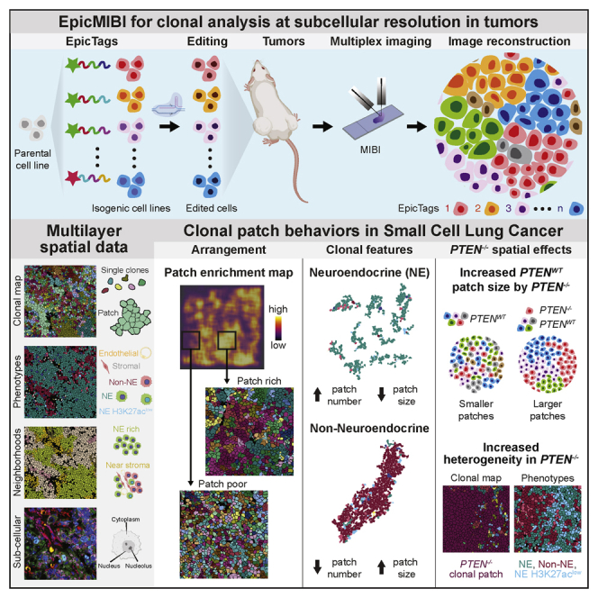
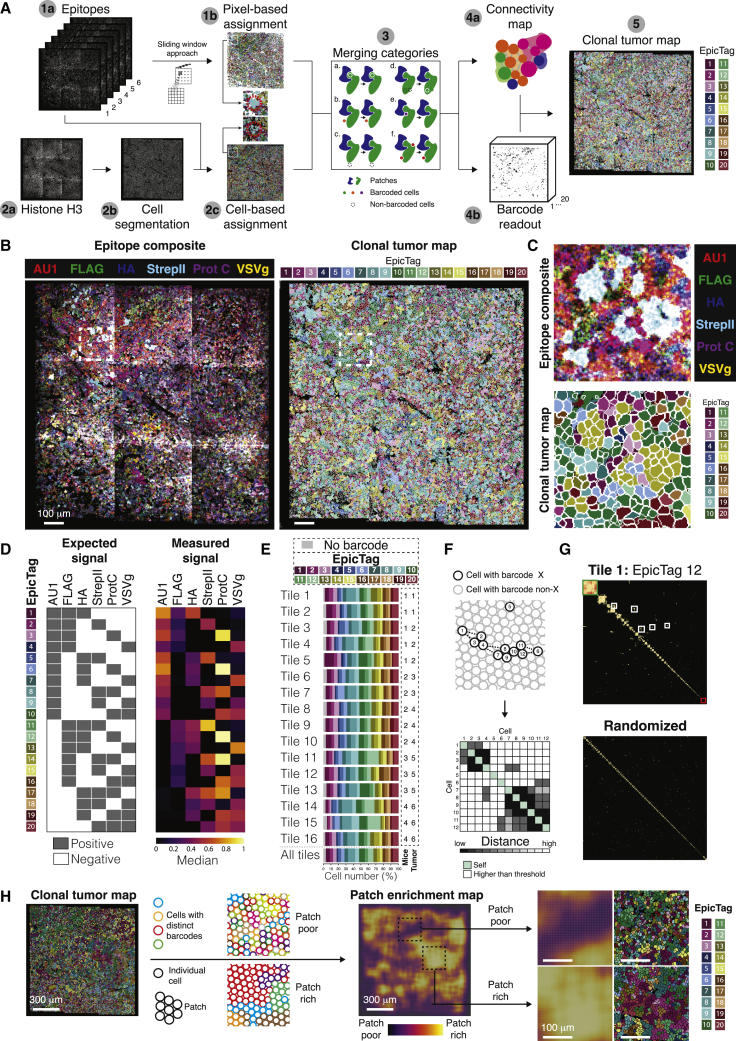
Intratumoral heterogeneity is a seminal feature of human tumors contributing to tumor progression and response to treatment. Current technologies are still largely unsuitable to accurately track phenotypes and clonal evolution within tumors, especially in response to genetic manipulations. Here, we developed epitopes for imaging using combinatorial tagging (EpicTags), which we coupled to multiplexed ion beam imaging (EpicMIBI) for in situ tracking of barcodes within tissue microenvironments. Using EpicMIBI, we dissected the spatial component of cell lineages and phenotypes in xenograft models of small cell lung cancer. We observed emergent properties from mixed clones leading to the preferential expansion of clonal patches for both neuroendocrine and non-neuroendocrine cancer cell states in these models. In a tumor model harboring a fraction of PTEN-deficient cancer cells, we observed a non-autonomous increase of clonal patch size in PTEN wild-type cancer cells. EpicMIBI facilitates in situ interrogation of cell-intrinsic and cell-extrinsic processes involved in intratumoral heterogeneity.

Keywords: MIBI; PTEN; SCLC; multiplex imaging; neuroendocrine; spatial barcoding; tumor heterogeneity.


Conflict of interest statement

Declaration of interests J.S. has equity in, and is an advisor for, DISCO Pharmaceuticals. G.P.N. and M.A. are co-founders and stockholders of Ionpath, Inc., and inventors on MIBI patents. The other authors declare no competing interests.

Figures

None
Graphical abstract
Figure 1
Figure 1
Imaging-based identification of specific cell populations with epitope-based barcodes (A) Schematic of a representative EpicTag construct. (B) Workflow for multiplex imaging of epitope-based barcodes in cell pellets. (C–E) Representative MIBI images of a cell pellet. (C) Left: overlay of anti-GFP and anti-histone H3 (HH3) images showing GFP+ (white arrow) and GFP cells (yellow arrow). Right: overlay of anti-GFP and the sum of all six anti-epitope images. Scale bars, 40 μm. (D) Representative anti-epitope images from the same field of view (FOV). The multicolor overlay at the bottom shows that each GFP+ cell expresses a combination of epitopes. Scale bars, 80 μm. (E) Multiple images showing a representative cell for each of the 20 barcodes. Dashed lines were manually drawn to indicate the contour of the relevant cell. The numbers within the white squares indicate whether the epitope was expected (1) or not (0). Images are enlargements from boxed regions in Figure S1G. Scale bars, 10 μm. See also Figure S1.
Figure 2
Figure 2
Detection of epitope-based barcodes in SCLC xenografts (A) Workflow for multiplex imaging of epitope-based barcodes in subcutaneous NCI-H82 SCLC xenografts. (B) Left: composite of anti-epitope images of a tumor consisting of a pooled population of 20 barcoded NCI-H82. Right: enlarged images from boxes in the left image showing that individual cells express three of the six epitopes. Scale bar, 80 μm. (C–E) Representative MIBI images of a tumor consisting of a pooled population of wild-type NCI-H82 cells and four barcoded NCI-H82 cell lines expressing GFP or mCherry tagged to six epitopes (AU1, FLAG, StrepII, Prot C, Tag100, and VSVg) or to three epitopes (FLAG, HA, and E2). (C) Top: schematic of a representative EpicTag construct variable on protein expression. Bottom left: overlay of anti-GFP, anti-mCherry, and anti-HH3 images showing cells expressing GFP or mCherry but not both simultaneously. Bottom right: enlarged images from the box on the left. Scale bar, 40 μm. (D) Top: schematic of a representative EpicTag construct variable on the number of epitopes. Bottom left: overlay of anti-E2, anti-Tag100, and anti-HH3 images showing cells expressing Tag100 or E2 but not both simultaneously. Bottom right: enlarged images from the box on the left. Scale bar, 40 μm. (E) Images of the same FOV showing cells expressing either GFP (green arrow) or mCherry (red arrow) and six epitopes. Scale bar, 8 μm. See also Figure S2.
Figure 3
Figure 3
Spatial organization of NCI-H82 SCLC xenografts (A) Summary of the antibody panel used for this study. (B) Images for each marker in a representative region of an NCI-H82 xenograft. HH3 is shown in all images (blue). Scale bars, 50 μm. (C) Images for αSMA, CD31, and human cell marker in a representative region of the dataset. Scale bars, 20 μm. (D) Images for 14 markers in a representative region of the dataset. These data exemplify antibody staining patterns used to assess barcode (left), phenotype (second to the left), epigenetic state (second to the right), and metabolic state (right). Scale bars, 100 μm (top) and 50 μm (bottom). (E) Images of γH2AX, Ki-67, and pS28 HH3 in a representative region of the dataset. Scale bars, 20 μm. (F) A heatmap of mean marker expression in the eight phenotypic clusters (rows). The intensity of each marker is defined by the color bar at the bottom (normalized counts). The mitotic phenotypic cluster is composed by non-NE and NE cells. (G) UMAP of all cells in the dataset (n = 231,715) colored by their phenotypic cluster. (H) Frequencies of each phenotypic cluster in the entire dataset (n = 231,715) and in each individual tile (tiles 1 to 16). (I) Representative phenotypic cluster maps on a region enriched in NE cells (tile 5; phenotypic cluster 7) and a region with distinct cell types (tile 15; phenotypic clusters 1 to 8). Scale bars, 100 μm. (J) Schematic of cellular neighborhoods. (K) A heatmap of the frequencies of each phenotypic cluster for each of the seven CNs (rows). (L) Representative neighborhood maps on a region enriched in homotypic NE cell interactions (tile 5; CN 3) and in a region with distinctive substructures (tile 15; CNs 1 to 7). Scale bars, 100 μm. See also Figure S3.
Figure 4
Figure 4
A debarcoding strategy identifies clonal tumor patches (A) Pipeline to reconstruct clonal tumor maps. (B and C) Representative clonal tumor map from a barcoded NCI-H82 xenograft. (B) Left: overlay of anti-epitope images. Right: clonal tumor map of the image on the left. Scale bars, 100 μm. (C) Enlarged view of the white boxes in (B). (D) Epitope signal on each debarcoded population. Left: expected signal for each epitope in each barcode. Gray indicates positive signal. Right: measured epitope signal in each debarcoded population from (B). Color shows the median of the normalized counts. (E) Frequencies of each EpicTag barcode in the dataset (n = 231,715) and in each individual tile (tiles 1 to 16). (F) Top: schematic representation of cells with the same EpicTag barcode. Cells with a particular barcode can be individually distributed (e.g., cell 1) or grouped (e.g., cells 2 to 4). Cells sharing a barcode can be distributed nearby (e.g., cells 6 and 11) or far apart (e.g., cell 5). Bottom: schematic grid of pairwise distances of the schematic shown on top. (G) Grid of pairwise interactions showing the distances of each cell to its fifth nearest neighbor for cells with EpicTag 12 in tile 1 (top) and a randomized sample (bottom). Randomization was performed by randomly assigning EpicTag 12 to segmented cells in tile 1, up to the number of EpicTag 12 in tile 1. Cells were arranged in the diagonal by patch size (larger patches in the top left corner). The green box exemplifies a clonal tumor patch. The white boxes exemplify clonal tumor patches that are closer in space. The red box exemplifies individually scattered cells. (H) A clonal tumor map (left image) can be decomposed based on the size of the clones into patch enrichment maps (middle image). Certain areas are enriched in patches (right, bottom image) or depleted (right, top image). See also Figure S4.
Figure 5
Figure 5
Non-neuroendocrine SCLC cells establish large patches with increased proliferative index and decreased DNA damage (A) Overview of the different layers obtained with epicMIBI. Scale bars, 100 μm. (B) Polar plots of marker expression in homotypic CNs (cluster 2, CN B; cluster 6, CN D; cluster 7, CN C). Synaptophysin (SYP) and citrate synthase (CS). (C) Total number of barcoded cells and patches for each EpicTag plotted for clusters 2 (non-NE) and 7 (NE) in all acquired tiles. (D) Representative image of large patches with phenotypic clusters and EpicTag barcodes indicated. White arrow highlights a large patch of non-NE cells. Yellow arrows indicate where non-NE and NE cells share the same barcode. Scale bars, 100 μm. (E) Patch sizes by cell number per EpicTag for cluster 2 (non-NE cells, red) and cluster 7 (NE cells, green). p = 2.210−16. (F–J) Single-cell analysis in individual clusters. For each cluster the data were separated by patch size: 1 cell, 1 to 10 cells, and over 10 cells. Significance is calculated by ANOVA within and between groups and adjusted by Bonferroni (Table S1). p-adj = 0.05–0.01, ∗∗p-adj = 0.01–0.001, ∗∗∗p-adj < 0.001. (F) Ki-67 expression. (G) Quiescent cells per cluster quantified by the percentage of cells per cluster that are negative for Ki-67 expression. (H) Citrate synthase expression. (I) GLUT1 expression. (J) Percentage of cells with DNA damage per cluster, inferred from γH2AX expression. See also Figure S5.
Figure 6
Figure 6
PTEN deficiency in a fraction of cancer cells modifies tumor architecture through cell-intrinsic and -extrinsic mechanisms (A) Workflow for multiplex imaging of genetically modified epitope-based barcoded cells in subcutaneous NCI-H82 xenografts. (B) UMAP of cultured cells (n = 69,065) grouped and colored by epitope expression. Dashed ellipses and percentages indicate PTEN−/− EpicTag 1 (bottom) and 20 (top). (C) Frequencies of each EpicTag barcode in cells grown in vitro and in vivo in “Control” and “PTEN−/−” pools. Black dashed boxes indicate PTEN−/− cells. (D) Frequencies of each phenotypic cluster in “Control” (n = 56,199) and “PTEN−/−” (n = 142,056) tumors. For the “PTEN−/−” tumors, frequencies for PTEN wild-type cells (“–”), PTEN unedited but nucleofected cells (“ctrl”), and PTEN knockout cells (“PTEN−/−”) are shown. (E) Frequencies of each CN in “Control” and “PTEN−/−” tumors. (F) Clonal tumor, phenotypic cluster, and CN maps, and epigenetic markers in a representative region of a PTEN−/− NCI-H82 xenograft highlighting wild-type cells. Bottom row images are enlarged representations of red dashed squares in the top row. White arrows indicate representative H3K27ac, H4K8ac+, and H3K4me2+ cells enriched in CN E. Scale bars, 40 μm (top) and 10 μm (bottom). (G) Overall patch size by cell number in “Control” and “PTEN−/−” tumors. The overall patch size from the dataset shown in Figure 5 is shown as reference (“First set control”). P values calculated by the Student’s t test. (H) Images of clonal tumor map with EpicTag barcodes highlighted in patches of more than 50 cells in “Control” and “PTEN−/−” tumors. White arrows indicate PTEN knockout patches. The legend indicates the color for each EpicTag barcode. The black square in the image at the bottom, second from the right, is consequence of a tile having eight FOVs instead of nine (see Figure S3C). Scale bars, 100 μm. (I) Patch size by cell number per EpicTag in “Control” and “PTEN−/−” tumors. P value calculated by the Student’s t test. (J) Tumor volume of 25 days of growth in mice. Groups: PTEN−/− cells (5 mice for replicate (Rep.) A, 6 mice for Rep. B), PTEN−/− 50%: wild-type (WT) 50% (5 mice for Rep. A, 6 mice for Rep. B), PTEN−/−10%: WT 90% (4 mice for Rep. A, 6 mice for Rep. B), WT 50%: WT 50% (5 mice for Rep. A, 6 mice for Rep. B). (K) Ratio of GFP to mCherry in NCI-H82 cells in vitro and in vivo at day 25. Mixes are 10:90 and 50:50 mixes for PTEN−/− (GFP):WT (mCherry) and 10:90 for WT (GFP):WT (mCherry). See also Figures S6 and S7.

References

    1. Angelo M., Bendall S.C., Finck R., Hale M.B., Hitzman C., Borowsky A.D., Levenson R.M., Lowe J.B., Liu S.D., Zhao S., et al. Multiplexed ion beam imaging of human breast tumors. Nat. Med. 2014;20:436–442. - PMC - PubMed
    1. Askary A., Sanchez-Guardado L., Linton J.M., Chadly D.M., Budde M.W., Cai L., Lois C., Elowitz M.B., Cai L., Lois C., Elowitz M.B. “In situ readout of DNA barcodes and single base edits facilitated by in vitro transcription. Nat. Biotechnol. 2020;38:66–75. - PMC - PubMed
    1. Bai Y., Zhu B., Rovira-Clave X., Chen H., Markovic M., Chan C.N., Su T.-H., McIlwain D.R., Estes J.D., Keren L., et al. Adjacent cell marker lateral spillover compensation and reinforcement for multiplexed images. Front. Immunol. 2021;12 - PMC - PubMed
    1. Baranski A., Milo I., Greenbaum S., Oliveria J.-P., Mrdjen D., Angelo M., Keren L. MAUI (MBI analysis user interface)-an image processing pipeline for multiplexed mass based imaging. PLoS Comput. Biol. 2021;17 - PMC - PubMed
    1. Boone P.G., Rochelle L.K., Ginzel J.D., Lubkov V., Roberts W.L., Nicholls P.J., Bock C., Flowers M.L., von Furstenberg R.J., Stripp B.R., et al. A cancer rainbow mouse for visualizing the functional genomics of oncogenic clonal expansion. Nat. Commun. 2019;10:5490. - PMC - PubMed

Publication types