. 2022 Dec 16;29(1):30-43.
doi: 10.1261/rna.079472.122.

A-MYB/TCFL5 regulatory architecture ensures the production of pachytene piRNAs in placental mammals

Affiliations

A-MYB/TCFL5 regulatory architecture ensures the production of pachytene piRNAs in placental mammals

Tiangxiong Yu et al. RNA. .

Abstract

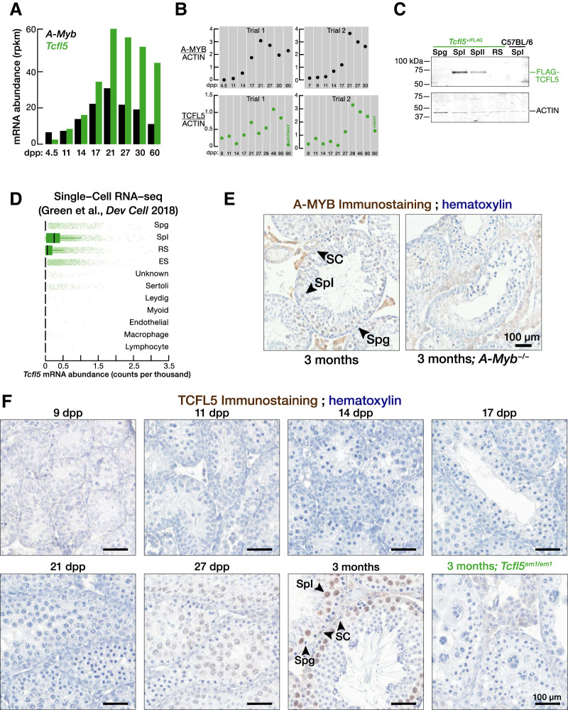
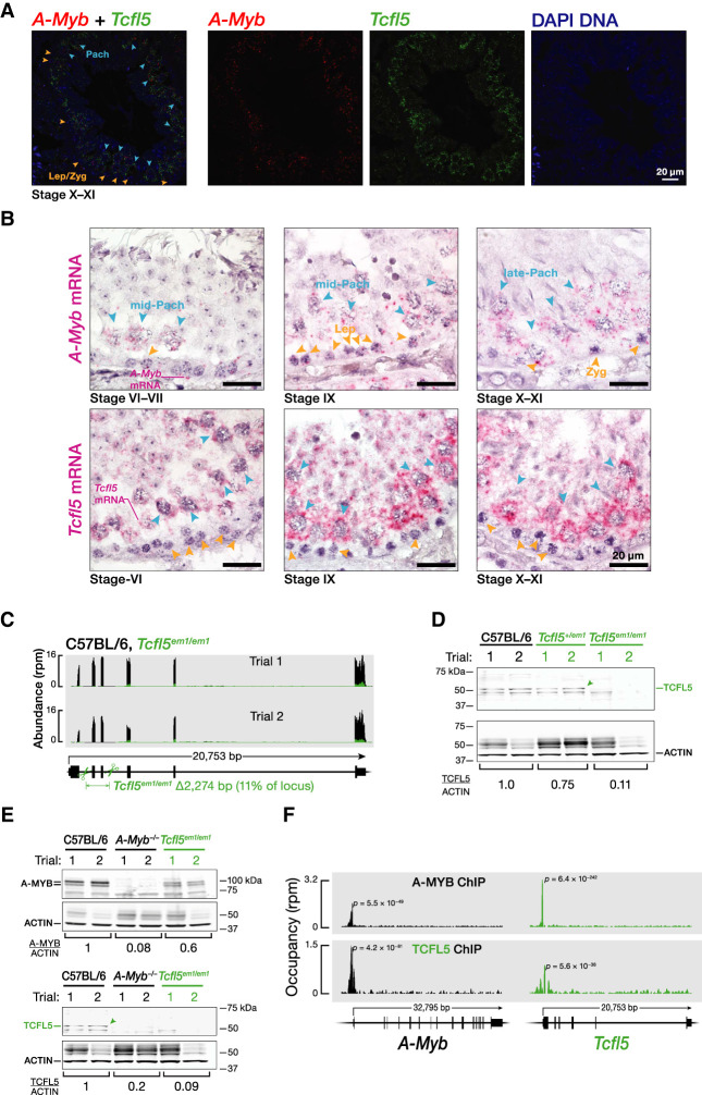
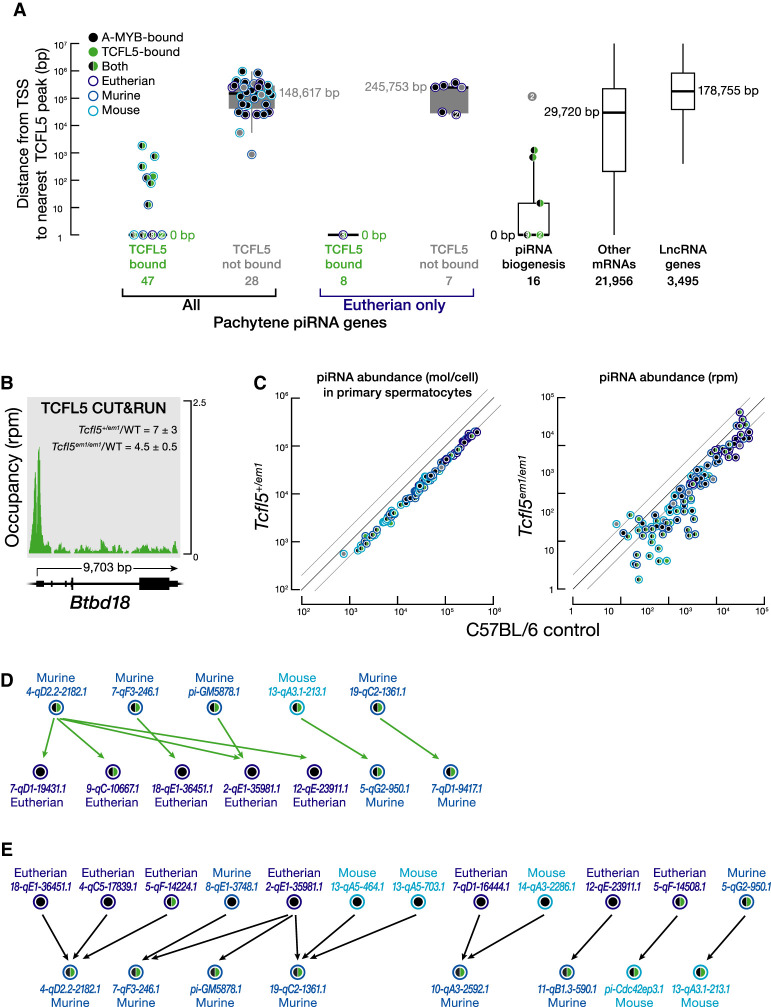
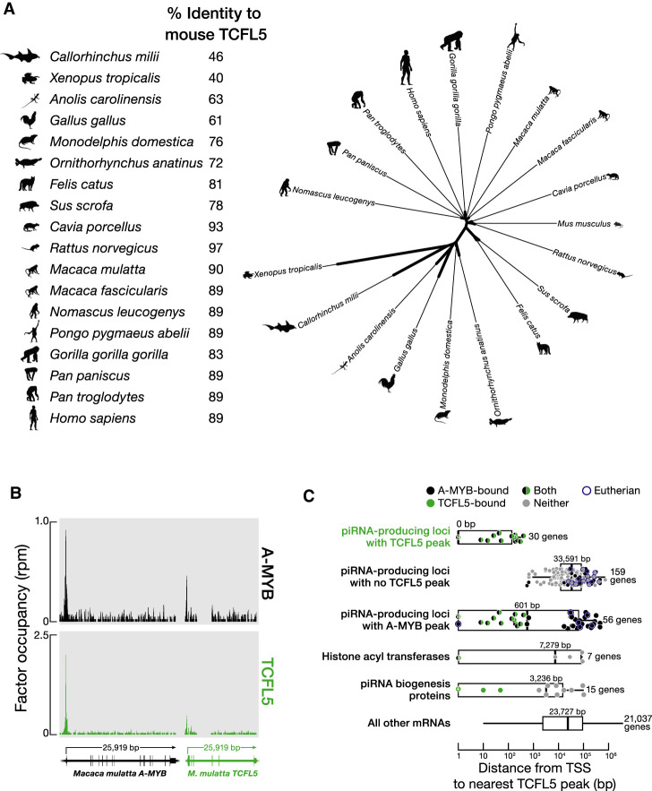
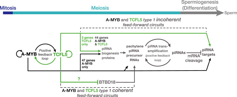
In male mice, the transcription factor A-MYB initiates the transcription of pachytene piRNA genes during meiosis. Here, we report that A-MYB activates the transcription factor Tcfl5 produced in pachytene spermatocytes. Subsequently, A-MYB and TCFL5 reciprocally reinforce their own transcription to establish a positive feedback circuit that triggers pachytene piRNA production. TCFL5 regulates the expression of genes required for piRNA maturation and promotes transcription of evolutionarily young pachytene piRNA genes, whereas A-MYB activates the transcription of older pachytene piRNA genes. Intriguingly, pachytene piRNAs from TCFL5-dependent young loci initiate the production of piRNAs from A-MYB-dependent older loci, ensuring the self-propagation of pachytene piRNAs. A-MYB and TCFL5 act via a set of incoherent feedforward loops that drive regulation of gene expression by pachytene piRNAs during spermatogenesis. This regulatory architecture is conserved in rhesus macaque, suggesting that it was present in the last common ancestor of placental mammals.

Keywords: A-MYB; TCFL5; pachytene piRNAs; spermatogenesis.


Figures

FIGURE 1.
FIGURE 1.
TCFL5 is expressed in pachytene spermatocytes during male meiosis I. (A,B) A-Myb and Tcfl5 mRNA (A) and protein abundance (B) at distinct developmental stages of mouse spermatogenesis. See Supplemental Figure S1A for uncropped western blot images. (C) Abundance of FLAG-TCFL5 protein in FACS-purified germ cell from Tcfl5+/FLAG mouse. Each lane contained protein from 75,000 germ cells. (D) mRNA abundance of Tcfl5 mRNA measured by single-cell sequencing by Green et al. (2018). (E) Immunohistochemical detection of A-MYB in adult testis section. A-Myb/ testis sections serve as antibody specificity controls. (F) Immunohistochemical detection of TCFL5 in testis sections from staged mice. Tcfl5em1/em1 testis sections serve as antibody specificity controls. (Spg) Spermatogonia, (SpI) primary spermatocytes, (SpII) secondary spermatocytes, (RS) round spermatids, (ES) elongating spermatids, (SC) Sertoli cells.
FIGURE 2.
FIGURE 2.
A-Myb expression precedes that of Tcfl5. (A,B) Temporal A-Myb and Tcfl5 mRNA expression in a seminiferous tubule section from adult mouse testis was detected. Two-color RNA fluorescent in situ hybridization (RNA-FISH) (A). Single-color RNA-FISH (B). (C) Strategy to generate Tcfl5 knockout mice using a single guide RNA (sgRNA) and Cas9. Scissors indicate sites targeted by sgRNAs designed to delete Tcfl5 exons 2 and 3. RNA-seq was used to measure the abundance of Tcfl5 mRNA in adult testes. (D) Abundance of TCFL5 protein in Tcfl5em1/em1 homozygous and Tcfl5+/em1 heterozygous mutant testes, relative to C57BL/6 wild-type testes. ACTIN serves as a loading control. Each lane contained 50 µg testis protein. (E) Abundance of A-MYB and TCFL5 proteins in A-Myb/ and Tcfl5em1/em1 homozygous mutant mouse testes. ACTIN serves as a loading control. Each lane contained 50 µg testis protein. (F) A-MYB and TCFL5 ChIP-seq peaks at the promoters of the A-Myb and Tcfl5 genes.
FIGURE 3.
FIGURE 3.
Younger pachytene piRNA genes whose transcription is activated by TCFL5 produce piRNAs that initiate piRNA production in evolutionarily older pachytene piRNA genes. (A) The distance from the nearest TCFL5 peak to the transcription start sites (TSS) of pachytene piRNA genes, genes encoding piRNA biogenesis proteins, and other protein-coding or noncoding genes obtained from CUT&RUN of FACS-purified primary spermatocytes. Horizontal lines: median; whiskers: maximum and minimum values, excluding outliers. Each dot represents distance (mean of two trials) from the nearest TCFL5 peak to the transcription start site of individual genes. Measurements with the same values are indicated by a single marker indicating the number of individual data points. Gpat2, which is required for piRNA biogenesis in flies (Vagin et al. 2013) and in cultured mouse germline stem cells (Shiromoto et al. 2013), was not included in this analysis, because its participation in the piRNA pathway in vivo in mice remains to be established. (B) TCFL5 CUT&RUN peak at the promoter of Btbd18. WT denotes C57BL/6. (C) Steady-state abundance of pachytene piRNAs in primary spermatocytes (left) or whole testes (right) for Tcfl5+/em1 heterozygotes compared to C57BL/6 controls. Each marker represents a pachytene piRNA gene. (D,E) Pachytene piRNA-directed cleavage sites in pachytene piRNA precursor transcripts. In D, each arrow points from the piRNAs derived from a TCFL5-dependent pachytene piRNA gene toward the corresponding cleavage site in a pachytene piRNA precursor transcript whose transcription is activated by A-MYB. TCFL5-dependent genes were defined as those whose promoters were occupied by TCFL5 and whose precursor and piRNA abundance in Tcfl5em1/em1 mutant mice were less than those in C57BL/6. In E, each arrow points from the piRNAs derived from one of 12 A-MYB-dependent pachytene piRNA genes toward genes for which the abundance of pachytene piRNA precursor transcripts was reduced less than twofold in Tcfl5em1/em1 mutant mice. A-MYB-dependent genes were defined as those whose promoters were occupied by A-MYB and whose expression was essentially unchanged in Tcfl5em1/em1 mutant mice.
FIGURE 4.
FIGURE 4.
The role of TCFL5 in piRNA production is conserved in rhesus macaque. (A, left) Percent identity of mouse TCFL5 to TCFL5 in other mammals. (Right) Unrooted tree of TCFL5 protein sequences, aligned using Clustal Omega. The unrooted tree was constructed using Randomized Axelerated Maximum Likelihood with default parameters and visualized in the Interactive Tree Of Life. (B) A-MYB and TCFL5 ChIP-seq peaks at the promoters of the rhesus macaque A-MYB and TCFL5 genes. (C) The distance from the nearest TCFL5 ChIP-seq peak to the transcription start site (TSS) for rhesus genes separated by class. Vertical lines: median; whiskers: maximum and minimum values, excluding outliers. A single marker containing the number of data points marks multiple identical values.
FIGURE 5.
FIGURE 5.
A model for the A-MYB/TCFL5 regulatory architecture of mouse male meiotic cells. The model incorporates hypotheses from Li et al. (2013), Özata et al. (2020), Wu et al. (2020), Choi et al. (2021), and this study. The figure highlights the roles of A-MYB and TCFL5 in regulating pachytene piRNA genes and direct targets of pachytene piRNAs during mouse spermatogenesis.

References

    1. Adiconis X, Borges-Rivera D, Satija R, DeLuca DS, Busby MA, Berlin AM, Sivachenko A, Thompson DA, Wysoker A, Fennell T, et al. 2013. Comparative analysis of RNA sequencing methods for degraded or low-input samples. Nat Methods 10: 623–629. 10.1038/nmeth.2483 - DOI - PMC - PubMed
    1. Alon U. 2007. Network motifs: theory and experimental approaches. Nat Rev Genet 8: 450–461. 10.1038/nrg2102 - DOI - PubMed
    1. Anders S, Pyl PT, Huber W. 2015. HTSeq—a Python framework to work with high-throughput sequencing data. Bioinformatics 31: 166–169. 10.1093/bioinformatics/btu638 - DOI - PMC - PubMed
    1. Aravin A, Gaidatzis D, Pfeffer S, Lagos-Quintana M, Landgraf P, Iovino N, Morris P, Brownstein MJ, Kuramochi-Miyagawa S, Nakano T, et al. 2006. A novel class of small RNAs bind to MILI protein in mouse testes. Nature 442: 203–207. 10.1038/nature04916 - DOI - PubMed
    1. Bastos H, Lassalle B, Chicheportiche A, Riou L, Testart J, Allemand I, Fouchet P. 2005. Flow cytometric characterization of viable meiotic and postmeiotic cells by Hoechst 33342 in mouse spermatogenesis. Cytometry A 65: 40–49. 10.1002/cyto.a.20129 - DOI - PubMed