. 2022 Oct 14;6(1):e202201449.
doi: 10.26508/lsa.202201449. Print 2023 Jan.

Low expression of EXOSC2 protects against clinical COVID-19 and impedes SARS-CoV-2 replication

Affiliations

Low expression of EXOSC2 protects against clinical COVID-19 and impedes SARS-CoV-2 replication

Tobias Moll et al. Life Sci Alliance. .

Abstract

New therapeutic targets are a valuable resource for treatment of SARS-CoV-2 viral infection. Genome-wide association studies have identified risk loci associated with COVID-19, but many loci are associated with comorbidities and are not specific to host-virus interactions. Here, we identify and experimentally validate a link between reduced expression of EXOSC2 and reduced SARS-CoV-2 replication. EXOSC2 was one of the 332 host proteins examined, all of which interact directly with SARS-CoV-2 proteins. Aggregating COVID-19 genome-wide association studies statistics for gene-specific eQTLs revealed an association between increased expression of EXOSC2 and higher risk of clinical COVID-19. EXOSC2 interacts with Nsp8 which forms part of the viral RNA polymerase. EXOSC2 is a component of the RNA exosome, and here, LC-MS/MS analysis of protein pulldowns demonstrated interaction between the SARS-CoV-2 RNA polymerase and most of the human RNA exosome components. CRISPR/Cas9 introduction of nonsense mutations within EXOSC2 in Calu-3 cells reduced EXOSC2 protein expression and impeded SARS-CoV-2 replication without impacting cellular viability. Targeted depletion of EXOSC2 may be a safe and effective strategy to protect against clinical COVID-19.


Conflict of interest statement

MP Snyder is a co-founder and member of the scientific advisory board of Personalis, Qbio, January, SensOmics, Protos, Mirvie, NiMo, Onza and Oralome. He is also on the scientific advisory board of Danaher, Genapsys and Jupiter. The Krogan Laboratory has received research support from Vir Biotechnology, F Hoffmann-La Roche, and Rezo Therapeutics. Nevan Krogan has financially compensated consulting agreements with the Icahn School of Medicine at Mount Sinai, New York, Maze Therapeutics, Interline Therapeutics, Rezo Therapeutics, GEn1E Lifesciences, Inc. and Twist Bioscience Corp. He is on the Board of Directors of Rezo Therapeutics and is a shareholder in Tenaya Therapeutics, Maze Therapeutics, Rezo Therapeutics, and Interline Therapeutics.

Figures
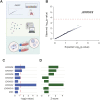
Figure 1.
Figure 1.. Unbiased screen of host proteins identified as high-confidence interacting partners of SARS-CoV-2 proteins links RNA exosome components to risk of clinical COVID-19.
(A) Schematic of the study design. Known host-viral interactions were screened for disease-association by combining lung-specific eQTLs with a genome-wide association studies for COVID-19 symptoms. Identification of a positive correlation between EXOSC2 expression and increased severity of COVID-19 led to further study of interactions between the SARS-CoV-2 polymerase and the entire human RNA exosome by AP-MS. Finally, CRISPR editing of EXOSC2 within human lung cells and subsequent infection with SARS-CoV-2 facilitated validation of the relationship between EXOSC2 expression and viral replication and interrogation of the underlying biological mechanism. (B) Lung eQTLs were used to group genetic variants according to their effect on expression of 332 host genes encoding proteins which interact with viral proteins. Only expression of EXOSC2 was significantly associated with clinical risk of COVID-19 after Bonferroni multiple testing (red line). (C, D) Lung eQTLs were used to group genetic variants according to their effect on expression of all genes encoding components of the RNA exosome. Expression levels of EXOSC7, EXOSC9, and EXOSC2 were significantly linked to clinical COVID-19, and in each case, higher expression was associated with higher risk of infection. P = 0.05 is indicated by a red dashed line.
Figure 2.
Figure 2.. AP-MS analysis confirms the interaction of the SARS-CoV-2 RNA polymerase with EXOSC2 and most of the components of the host RNA exosome.
Replicate affinity purifications of HEK293T cells expressing Strep-Nsp8 and untagged Nsp7 and control purifications (mock-transfected) were analysed by label-free quantitative mass spectrometry. Volcano plot of Strep-Nsp8 pulldowns from cells co-expressing Nsp7 compared with mock-transfected cells. RNA exosome complex proteins within the set of enriched proteins are labelled.
Figure S1.
Figure S1.. CRISPR editing with sgRNA targeted against EXOSC2 in Calu-3 cells.
(A) Sanger sequencing traces demonstrating spCas9 cut site adjacent to PAM and subsequent waveform decomposition in EXOSC2 edited cells. (B) Indel distribution of EXOSC2 edited Calu-3 cells. (C) qPCR reveals that expression of EXOSC2 mRNA is reduced in edited Calu-3 cells; comparison is made with control CRISPR editing targeting HPRT. (D) Immunoblotting for EXOSC2 protein revealed reduced expression in EXOSC2 edited Calu-3 cells compared with HPRT edited cells and WT unedited cells; recovery of EXOSC2 expression was achieved with EXOSC2 reconstitution. GAPDH served as a loading control, and a serial dilution of protein from WT cells is shown for reference.
Figure 3.
Figure 3.. Reduced expression of EXOSC2 in Calu-3 cells is not toxic and leads to reduced viral replication.
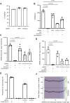
(A) Calu-3 cells were targeted with the indicated sgRNAs and cell viability was analysed by MTT assay. Data for unedited control cells were set to 100%. (B, C, D, E, F) Calu-3 cells targeted with sgRNAs and subsequently reconstituted with EXOSC2 as indicated were infected with SARS-CoV-2 (MOI = 1) for 17 h. As a negative control, cells infected with virus were exposed to a neutralising antibody. (B) Viral titres in supernatant samples were analysed by TCID50 assay. (C, D) Viral RNA levels were measured by absolute RT-qPCR quantification of N1 and N2 SARS-CoV-2 genomic RNA. (E) Viral genomic reads as a proportion of total RNA sequencing reads. (F) Viral genomic RNA sequencing reads mapped across the SARS-CoV-2 genome by normalised read-depth; colours represent distinct viral transcripts. Data are from three independent biological repeats. In panels (A, B, C, D, E), individual data points are shown with mean and SE. Significance was tested by the paired t test, and P-values are indicated.
Figure S2.
Figure S2.. Immunoblotting for RNA exosome components in Calu-3 cells with reduced EXOSC2 expression.
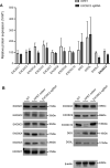
Immunoblotting for the total set of RNA exosome components within Calu-3 cells where CRISPR editing has been performed with sgRNA targeted against EXOSC2 to reduce EXOSC2 expression. (A, B) No other RNA exosome component is significantly depleted in three biological repeats (A); an example immunoblot is shown (B).
Figure 4.
Figure 4.. Transcriptomic analysis confirmed the inflammatory response to SARS-CoV-2 infection of Calu-3 cells and identified up-regulation of OAS genes in the context of reduced EXOSC2 expression.
RNA for sequencing was extracted from Calu-3 cells in the presence and absence of CRISPR editing with sgRNA targeted against EXOSC2; with and without infection with SARS-CoV-2 (MOI = 1) at 17 h; three biological replicates were obtained for all conditions. (A) First and second principal components for total gene expression across all sequenced samples. Samples include WT unedited Calu-3 cells and EXOSC2 edited Calu-3 cells; ± indicates the presence/absence of SARS-CoV-2 infection. (B) Heatmap representation of genes up-regulated in WT cells in the presence of SARS-CoV-2 infection. A darker colour indicates higher expression. (C) Volcano plot to compare gene expression in uninfected Calu-3 cells with and without CRISPR editing of EXOSC2. Dotted lines represent fold change of ± 2 and a Bonferroni multiple testing threshold for P-value by the genewise exact test. (D) Heatmap representation of 397 interferon-stimulated genes (Schoggins et al, 2011) across all sequenced samples. (D, E, F) Normalised expression of OAS1 (D) and OAS3 (E) in all four conditions.
Figure S3.
Figure S3.. Transcriptomic analysis of the effect of SARS-CoV-2 infection and/or reduced EXOSC2 expression in Calu-3 cells.
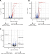
(A, B) Volcano plot to compare gene expression changes coincident with SARS-CoV-2 infection in (A) WT unedited Calu-3 cells and (B) in cells with prior CRISPR editing of EXOSC2. (C) Volcano plot to compare gene expression in SARS-CoV-2 infected Calu-3 cells in the presence and absence of CRISPR editing of EXOSC2. Dotted lines represent fold change of ± 2 and a Bonferroni multiple testing threshold for P-value by the genewise exact test.

Update of

References

    1. Allmang C, Mitchell P, Petfalski E, Tollervey D (2000) Degradation of ribosomal RNA precursors by the exosome. Nucleic Acids Res 28: 1684–1691. 10.1093/nar/28.8.1684 - DOI - PMC - PubMed
    1. Bray NL, Pimentel H, Melsted P, Pachter L (2016) Near-optimal probabilistic RNA-seq quantification. Nat Biotechnol 34: 525–527. 10.1038/nbt.3519 - DOI - PubMed
    1. Brodin P (2021) Immune determinants of COVID-19 disease presentation and severity. Nat Med 27: 28–33. 10.1038/s41591-020-01202-8 - DOI - PubMed
    1. Cagno V (2020) SARS-CoV-2 cellular tropism. Lancet Microbe 1: e2–e3. 10.1016/s2666-5247(20)30008-2 - DOI - PMC - PubMed
    1. Choi UY, Kang J-S, Hwang YS, Kim Y-J (2015) Oligoadenylate synthase-like (OASL) proteins: Dual functions and associations with diseases. Exp Mol Med 47: e144. 10.1038/emm.2014.110 - DOI - PMC - PubMed

Publication types

MeSH terms

Associated data