. 2022 Oct 14;15(1):146.
doi: 10.1186/s13045-022-01367-4.

PARP1-MGMT complex underpins pathway crosstalk in O6-methylguanine repair

Affiliations

PARP1-MGMT complex underpins pathway crosstalk in O6-methylguanine repair

Jodie D Cropper et al. J Hematol Oncol. .

Abstract

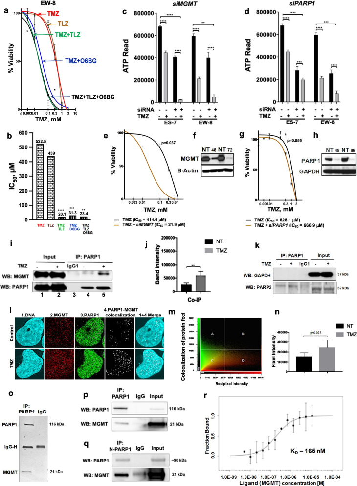
DNA lesions induced by alkylating agents are repaired by two canonical mechanisms, base excision repair dependent on poly(ADP) ribose polymerase 1 (PARP1) and the other mediated by O6-methylguanine (O6meG)-DNA methyltransferase (MGMT) in a single-step catalysis of alkyl-group removal. O6meG is the most cytotoxic and mutagenic lesion among the methyl adducts induced by alkylating agents. Although it can accomplish the dealkylation reaction all by itself as a single protein without associating with other repair proteins, evidence is accumulating that MGMT can form complexes with repair proteins and is highly regulated by a variety of post-translational modifications, such as phosphorylation, ubiquitination, and others. Here, we show that PARP1 and MGMT proteins interact directly in a non-catalytic manner, that MGMT is subject to PARylation by PARP1 after DNA damage, and that the O6meG repair is enhanced upon MGMT PARylation. We provide the first evidence for the direct DNA-independent PARP1-MGMT interaction. Further, PARP1 and MGMT proteins also interact via PARylation of MGMT leading to formation of a novel DNA damage inducible PARP1-MGMT protein complex. This catalytic interaction activates O6meG repair underpinning the functional crosstalk between base excision and MGMT-mediated DNA repair mechanisms. Furthermore, clinically relevant 'chronic' temozolomide exposure induced PARylation of MGMT and increased binding of PARP1 and MGMT to chromatin in cells. Thus, we provide the first mechanistic description of physical interaction between PARP1 and MGMT and their functional cooperation through PARylation for activation of O6meG repair. Hence, the PARP1-MGMT protein complex could be targeted for the development of advanced and more effective cancer therapeutics, particularly for cancers sensitive to PARP1 and MGMT inhibition.

Keywords: Cancer therapy; DNA damage and repair; Ewing sarcoma; MGMT; O6-Methylguanine; PARP1; Protein interaction.


Conflict of interest statement

The authors declare that they have no competing interests.

Figures

Fig. 1
Fig. 1
Pharmacological and genetic inhibition of PARP1 and MGMT potentiates temozolomide cytotoxicity in a linear fashion and is associated with PARP1-MGMT interaction. a TMZ-treated (0–3 mM) Ewing sarcoma cell lines exposed to TLZ (IC10) and O6BG (5 μM) for 96 h (Alamar Blue assay). EW-8 cell line is shown as a model example, additional results for ES-4, ES-6, and ES-7 cell lines are available in Additional File 1: Fig. S1a. TLZ, talazoparib. TMZ, temozolomide. O6BG, O6-benzylguanine. b Potentiation to TMZ: IC50 values for EW-8 cell line as in a. EW-8 cells are intrinsically resistant to TLZ [8]. P-values are calculated for TMZ vs TMZ + TLZ, TMZ + O6BG, TMZ + TLZ + O6BG by ANOVA3 followed by Tukey’s test for multiple comparisons: ****p ≤ 0.0001; ***p ≤ 0.001; **p ≤ 0.01; *p ≤ 0.05. Legend colors are coordinated with colors in a. c MGMT and d PARP1 gene knockdown-induced potentiation to TMZ (IC50, 48 h) in ES-7 and EW-8 cells (RNAi high-throughput screen). Readout is ATPlite cell viability assay. Each bar represents mean IC50; error bars are calculated for 3 siRNAs run in triplicate. P-values calculated by t-test, non-paired, un-equal variance, 2-sided: ****p ≤ 0.0001; ***p ≤ 0.001. PARP1 gene knockdown was not as effective as talazoparib, which inhibits PARP1 and PARP2 (the latter is linked to toxicity [9]). e TMZ treatment of EW-8 cells (0–1 mM) ± MGMT or g PARP1 gene knockdown by siRNA (Alamar Blue staining). Student’s paired 2-tailed t-test: p = 0.05 (e); *p ≤ 0.05 (g). f MGMT or h PARP1 protein downregulation by siRNA (Western blot at 48, 72, 96 h). GAPDH (37 kDa). Beta-actin (43 kDa). NT, no treatment. i EW-8 cells ± TMZ treatment (1 mM, 2 h): PARP1 pulldown was followed by PARP1 (top) or MGMT (bottom) immunoblotting. Lanes 1–2: co-immunoprecipitation. Lane 3: IgG1. Lanes 4–5: input. j Mean of protein band intensities generated from 3 independent co-immunoprecipitation experiments in (i). Student’s paired 2-tailed t-test: *p ≤ 0.05; **p ≤ 0.01 (see reverse co-IP in Additional File 1: Fig. S2c, d). k Negative (PARP1-GAPDH) and positive (PARP1-PARP2) interactions by co-immunoprecipitation in EW-8 cells. Samples prepared as in (i). IgG control is in middle lane. l Representative image of EW-8 cells nuclei staining with Hoechst 33,342 (blue, nuclei), Alexa Fluor 647 (red, MGMT), and Alexa Fluor 488 (green, PARP1). White pixels indicate green and red overlap, i.e., co-localization of PARP1 and MGMT. Top panel, no TMZ. Bottom panel, TMZ at 1 mM for 2 h. m Scatterplot representing red (MGMT) and green (PARP1) pixel intensities in (l); overlap of these colors along the diagonal in the field ‘c’ (~ 45°) corresponds to protein co-localization dots (shown as white pixels). Co-localization analysis was done using CellSens software (v2.1). Images were developed with Fluoview FV3000. n Quantification of white-pixel number of co-localized PARP1-MGMT sites in control vs TMZ-treated EW-8 cell nuclei. Data from 3 independent experiments were used for the analysis. NT, no treatment. o SDS-PAGE and silver staining of protein gel showing PARP1-MGMT interaction by immunoprecipitation assay. Pulldown with full-length PARP1. p Purified PARP1 and MGMT protein interaction. Mixed full-length PARP1 and MGMT proteins (1:1) were subjected to co-immunoprecipitation. PARP1 was pulled down with the co-immunoprecipitation specific PARP1 antibody (cst-9532) and immunoblotted with PARP1 (top; cst-9542) or MGMT (bottom; sc-241154) antibodies. IgG control is in the middle lane. q Purified N-terminal of PARP1 (aa 1–662) was mixed with full-length MGMT (1:1) and processed for co-immunoprecipitation. Samples prepared as in (p). IgG control shown in middle lane. r An MST-on time of 10 s analysis of the full-length PARP1 and MGMT protein affinity was performed using Monolith NT.115 at 17% LED power and medium MST power
Fig. 2
Fig. 2
Alkylating DNA damage intensifies O6meG repair through PARylation of MGMT. a SDS-PAGE/Western blot (top and middle) and BSA Ponceau staining (bottom) for PAR and MGMT. Key: Ss/dsOligo1 is MCAT; ss/dsOligo2 is MGMT-Oligo; ss/dsOligo3 is ss/dsMGMT-O6meG. The resulting proteins were detected by SDS-PAGE analysis followed by Western blot for PAR (top) and MGMT (bottom). See PARP1 Western blot in Additional File 1: Fig. S3b. BSA, Ponceau S membrane staining. PAR-PARP1 is auto-PARylated PARP1. PAR-MGMT is PARylated MGMT. b Glutathione-S-transferase (GST) is a non-binding substrate of PARP1 and is not PARylated (serves as control). The purified PARP1, GST proteins, NAD+, and dsOligo1 were processed as in (a). c ELISA assay to evaluate PAR levels in Ewing sarcoma EW-8, rhabdomyosarcoma (RD), and fibroblast HFF1 cell lines ± temozolomide treatment (1 mM, 2 h) using SpectraMax M5 plate reader (450 nm). Student’s paired 2-tailed t-test: **p ≤ 0.01. NT, no treatment. TMZ, temozolomide. d MGMT repair assay diagram. The repair product is cleavable by PvuII restriction digestion. The unrepaired O6meG dsDNA (intact, i.) and repair product (cleaved, c.) can be analyzed by gel electrophoresis. e MGMT repair assay. MGMT and PARP1 (6.2 nM) were incubated with MCAT dsDNA for 1 h at 37°C to induce PARylation and then incubated with 32P-labeled-O6meG-dsDNA (50 nM) for repair reaction. Reaction products were analyzed by PvuII treatment followed by PAGE and phosphor-imaging. f % of repair results quantified using Image J as a ratio of cleaved band intensity to a sum of intact and cleaved band intensities from (e) were plotted (by Prism 8). Stronger increase in DNA cleavage (O6meG repair) was observed in the presence of PARP1 and NAD+. Student’s paired 2-tailed t-test: **p ≤ 0.01. g PARylation activity in EW-8 cells in response to short- (2 mM, 2 h) and long-term (100 μM, 72 h) temozolomide treatment by Western blot for PAR, PARP1, MGMT, and GAPDH proteins. PAR-PARP1 is PARylated PARP1. h Quantified band intensities for PAR, PARP1, and MGMT bands normalized to GAPDH levels (n = 3) and plotted using Image J. GAPDH (37 kDa) is loading control. i Chromatin and nuclear soluble fractions of EW-8 cells treated with temozolomide at 2 mM for 2 h or at 100 μM for 72 h by Western blot. Histone 3 (15 kDa) is chromatin fraction control. SP1 (81 kDa) is nuclear soluble fraction control

References

    1. Gill SJ, Travers J, Pshenichnaya I, Kogera FA, Barthorpe S, Mironenko T, et al. Combinations of PARP Inhibitors with temozolomide drive PARP1 trapping and apoptosis in Ewing's Sarcoma. PLoS ONE. 2015;10(10):e0140988. doi: 10.1371/journal.pone.0140988. - DOI - PMC - PubMed
    1. Lord CJ, Ashworth A. PARP inhibitors: synthetic lethality in the clinic. Science. 2017;355(6330):1152–1158. doi: 10.1126/science.aam7344. - DOI - PMC - PubMed
    1. Smith MA, Reynolds CP, Kang MH, Kolb EA, Gorlick R, Carol H, et al. Synergistic activity of PARP inhibition by talazoparib (BMN 673) with temozolomide in pediatric cancer models in the pediatric preclinical testing program. Clin Cancer Res. 2015;21(4):819–832. doi: 10.1158/1078-0432.CCR-14-2572. - DOI - PMC - PubMed
    1. Schafer ES, Rau RE, Berg SL, Liu X, Minard CG, Bishop AJR, et al. Phase 1/2 trial of talazoparib in combination with temozolomide in children and adolescents with refractory/recurrent solid tumors including Ewing sarcoma: a children's oncology group phase 1 consortium study (ADVL1411) Pediatr Blood Cancer. 2020;67(2):e28073. doi: 10.1002/pbc.28073. - DOI - PMC - PubMed
    1. Smith MA, Hampton OA, Reynolds CP, Kang MH, Maris JM, Gorlick R, et al. Initial testing (stage 1) of the PARP inhibitor BMN 673 by the pediatric preclinical testing program: PALB2 mutation predicts exceptional in vivo response to BMN 673. Pediatr Blood Cancer. 2015;62(1):91–98. doi: 10.1002/pbc.25201. - DOI - PMC - PubMed

Publication types

MeSH terms