. 2022 Oct 13;14(19):7972-7985.
doi: 10.18632/aging.204326. Epub 2022 Oct 13.

DAZAP1 facilitates the alternative splicing of KITLG to promote multiple myeloma cell proliferation via ERK signaling pathway

Affiliations

DAZAP1 facilitates the alternative splicing of KITLG to promote multiple myeloma cell proliferation via ERK signaling pathway

Yanyan Zhou et al. Aging (Albany NY). .

Abstract

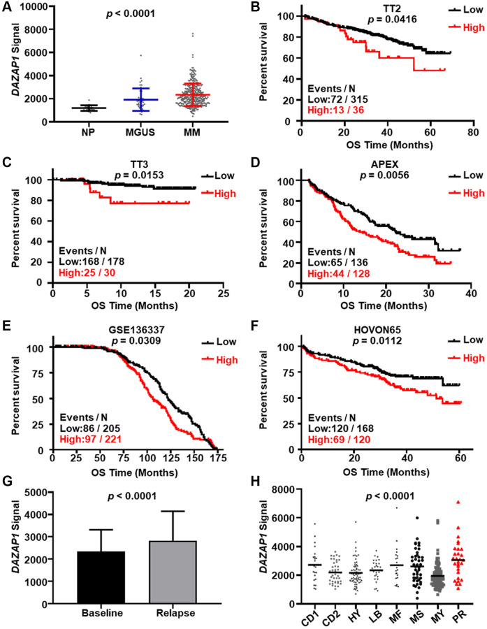
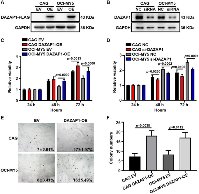
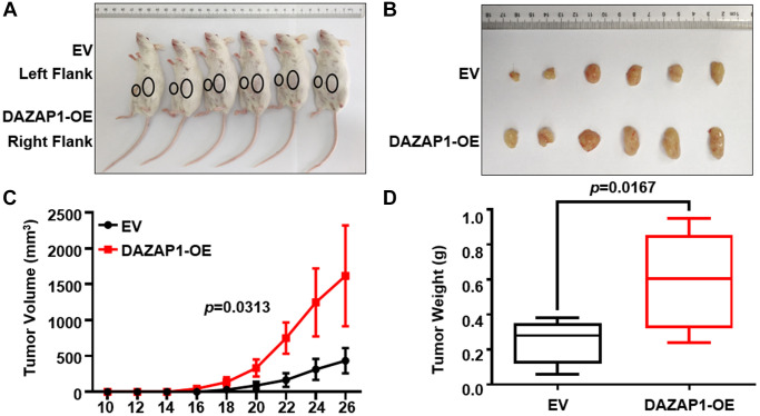
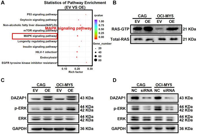
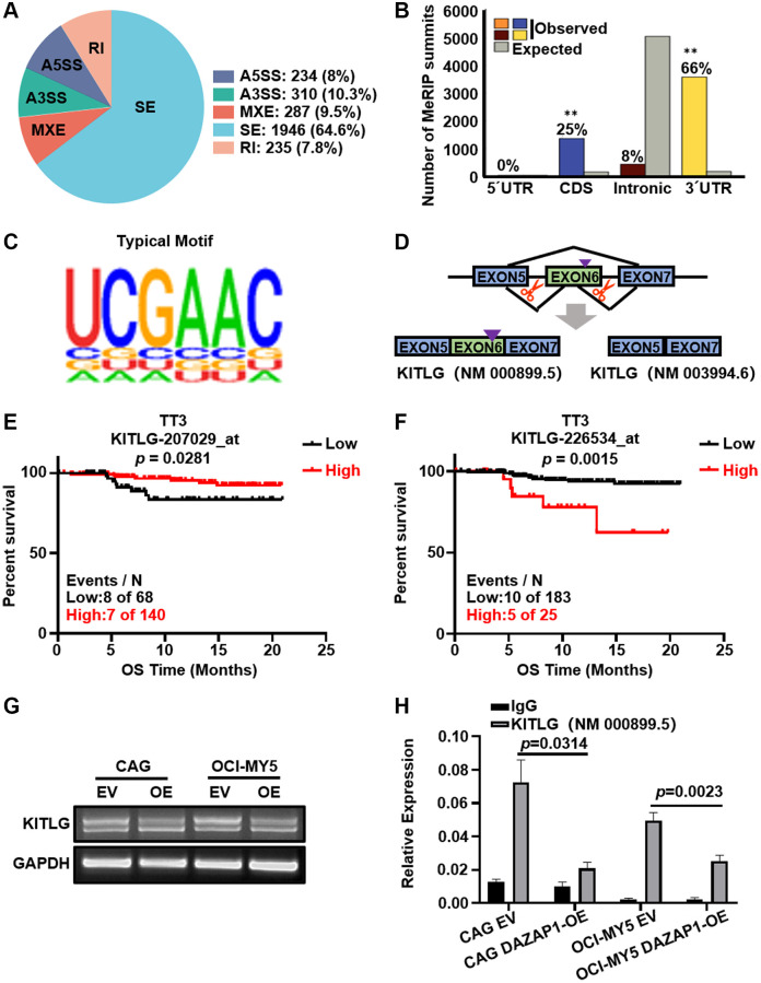
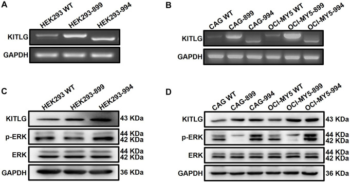
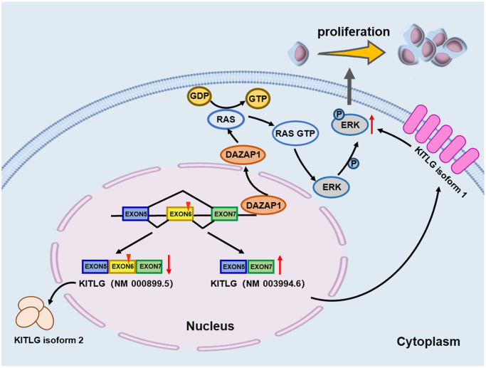
Multiple myeloma (MM) is an incurable plasma cell malignancy, in which alternative pre-mRNA splicing (AS) acts as one of the key transcriptome modifier. The Deleted in Azoospermia-Associated Protein 1 (DAZAP1) is a splicing factor that has been identified as an oncogene in multiple cancers, yet its role in MM proliferation remains unclear. We first analyzed MM clinical databases and found that MM patients with elevated DAZAP1 had a poor survival. Furthermore, we overexpressed DAZAP1 by lentiviral transfection and utilized siRNA silencing the expression of DAZAP1 in MM cells. DAZAP1 promoted MM cell proliferation in vitro and accelerated MM xenograft tumor growth in vivo. KEGG pathway enrichment analysis showed that ERK signaling pathway was activated in DAZAP1-OE MM cells. The analyses of RIP-seq and RIP-qPCR revealed that DAZAP1 activated alternative splicing of KIT proto-oncogene ligand (KITLG) mRNA. Further study validated that DAZAP1 increased ERK phosphorylation via modulating alternative splicing of KITLG mRNA to promote MM cell proliferation. In conclusion, we establish DAZAP1 as a tumor-promoting gene with therapeutic potential and provide mechanistic insights into targeting DAZAP1 as a new strategy for the diagnosis and treatment of MM.

Keywords: DAZAP1; ERK; KITLG; alternative splicing; multiple myeloma.


Conflict of interest statement

CONFLICTS OF INTEREST: The authors declare no conflicts of interest related to this study.

Figures

Figure 1
Figure 1
Elevated DAZAP1 is associated with poor outcome of MM patients. (A) DAZAP1 expression in different stages of MM from the GSE5900 dataset as shown in the graph. The signal level of DAZAP1 (226620_at signaling) was shown on the y-axis. The groups of normal plasma (NP, n = 22), monoclonal gammopathy of undetermined significance (MGUS, n = 44), and multiple myeloma (MM, n = 351) were sorted on the x-axis, respectively. (BF) Kaplan-Meier analysis of overall survival (OS) divided by high and low DAZAP1 expression in TT2, TT3, APEX, HOVON65 and GSE136337 cohorts. The number of patients in the cohorts was 351, 208, 264, 426 and 288, respectively. (G) DAZAP1 expression was increased in relapsed patient samples relative to the first diagnosis samples. (H) A box-plot showed DAZAP1 expression in eight MM subgroups.
Figure 2
Figure 2
Overexpression of DAZAP1 enhances the proliferative capacity of MM cells in vitro. (A and B) DAZAP1 expression in DAZAP1-OE and siDAZAP1 MM cells were examined by WB analysis. (C and D) The proliferation rate of DAZAP1-OE and siDAZAP1 MM cells was assessed by CCK8 assay. (E) Soft agar colony formation assay revealed overexpressed DAZAP1 accelerating colony formation. (F) The histogram showed quantification of colony formation in soft agar. All data management and analysis were done using the GraphPad Prism 8.0 version. The P value was calculated with Student’s t-test, (*p < 0.05, **p < 0.01, ***p < 0.001).
Figure 3
Figure 3
Increased DAZAP1 is conducive to tumor growth in MM xenograft model in vivo. (A) Photographic images of tumor burden mice were captured on day 26. (B) Subcutaneous tumors were gathered on the 26th day post grafting. (C) Growth curve of transplanted tumors. (D) Quantification of tumors weight from dissected tumors. All data are displayed as mean ± SD (*p < 0.05, **p < 0.01, ***p < 0.001).
Figure 4
Figure 4
DAZAP1 promotes the phosphorylation of ERK in MM cells. (A) A bubble diagram of the top 10 KEGG pathways. In the bubble diagram, the vertical axis indicates the KEGG pathways and the horizontal axis represents the enrichment ratio. The sizes of the dots indicate the number of genes in the Gene Ontology term. (B) Active RAS Pull-down assay and Western blot showed the levels of active form RAS protein compared to total RAS protein. (C and D) WB test examined the phosphorylation level of ERK expression in DAZAP1-OE and siDAZAP1 MM cells.
Figure 5
Figure 5
DAZAP1 triggers alternative splicing of KITLG mRNA in MM cells. (A) AS events were classified into five categories: skipped exon (SE), alternative 5′ splice site (A5SS), alternative 3′ splice site (A3SS), mutually exclusive exon (MXE), and retained intron (RI). (B) Binding of DAZAP1 was close to the 3′ splice site. (C) The typical motif on the peak-bound mRNA regions. (D) Schematic diagrams showed the alternative splicing of KITLG. (E and F) Two different probes of KITLG corresponded to different patient survivals in TT3 cohort. (G and H) RNA levels of different isoform of KITLG were tested by PCR and qPCR assays.
Figure 6
Figure 6
Abnormal alternative splicing of KITLG activates the ERK signaling pathway. (A and B) Agarose gel electrophoresis demonstrated the transfer efficiency on different isoforms of KITLG in both HEK293 and MM cells. (C and D) WB test verified that phosphorylated ERK expression was enhanced in KITLG isoform1-OE cells and downregulated in KITLG isoform2-OE cells.
Figure 7
Figure 7
Schematic depiction illustrates that DAZAP1 promotes MM cell proliferation through alternative splicing of KITLG.

Similar articles

Cited by

References

    1. Kazandjian D. Multiple myeloma epidemiology and survival: A unique malignancy. Semin Oncol. 2016; 43:676–81. 10.1053/j.seminoncol.2016.11.004 - DOI - PMC - PubMed
    1. Gupta N, Sharma A, Sharma A. Emerging biomarkers in Multiple Myeloma: A review. Clin Chim Acta. 2020; 503:45–53. 10.1016/j.cca.2019.12.026 - DOI - PubMed
    1. Röllig C, Knop S, Bornhäuser M. Multiple myeloma. Lancet. 2015; 385:2197–208. 10.1016/S0140-6736(14)60493-1 - DOI - PubMed
    1. Lê GN, Bones J, Coyne M, Bazou D, Dowling P, O'Gorman P, Larkin AM. Current and future biomarkers for risk-stratification and treatment personalisation in multiple myeloma. Mol Omics. 2019; 15:7–20. 10.1039/c8mo00193f - DOI - PubMed
    1. Joshua DE, Bryant C, Dix C, Gibson J, Ho J. Biology and therapy of multiple myeloma. Med J Aust. 2019; 210:375–80. 10.5694/mja2.50129 - DOI - PubMed

Publication types