. 2022 Oct 15;22(1):1068.
doi: 10.1186/s12885-022-10155-9.

Pan-cancer analysis of oncogenic TNFAIP2 identifying its prognostic value and immunological function in acute myeloid leukemia

Affiliations

Pan-cancer analysis of oncogenic TNFAIP2 identifying its prognostic value and immunological function in acute myeloid leukemia

Mei-Si Lin et al. BMC Cancer. .

Abstract

Background: Tumor necrosis factor alpha-induced protein 2 (TNFAIP2), a TNFα-inducible gene, appears to participate in inflammation, immune response, hematopoiesis, and carcinogenesis. However, the potential role of TNFAIP2 in the development of acute myeloid leukemia (AML) remains unknow yet. Therefore, we aimed to study the biological role of TNFAIP2 in leukemogenesis.

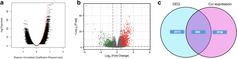
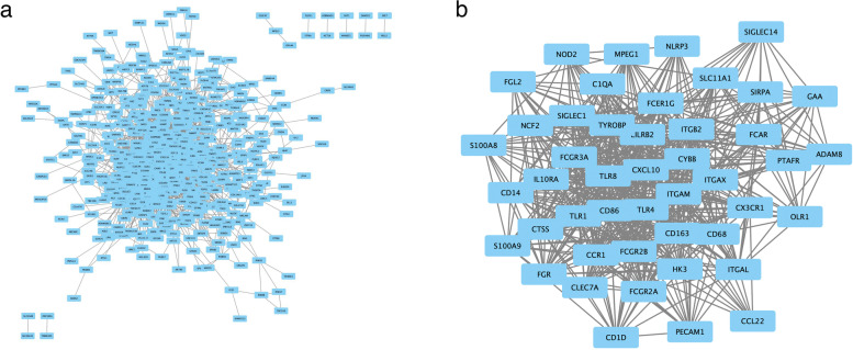
Methods: TNFAIP2 mRNA level, prognostic value, co-expressed genes, differentially expressed genes, DNA methylation, and functional enrichment analysis in AML patients were explored via multiple public databases, including UALCAN, GTEx portal, Timer 2.0, LinkedOmics, SMART, MethSurv, Metascape, GSEA and String databases. Data from The Cancer Genome Atlas (TCGA), Gene Expression Omnibus (GEO) and Beat AML database were used to determine the associations between TNFAIP2 expression and various clinical or genetic parameters of AML patients. Moreover, the biological functions of TNFAIP2 in AML were investigated through in vitro experiments.

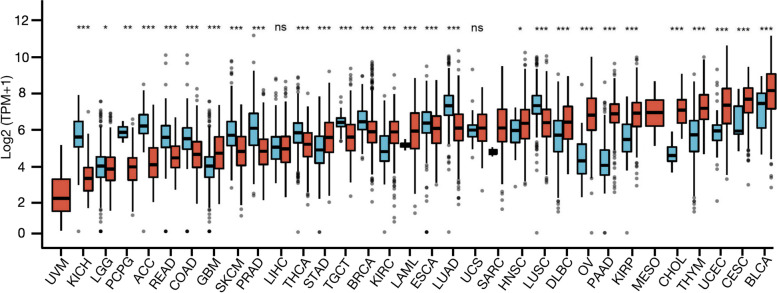
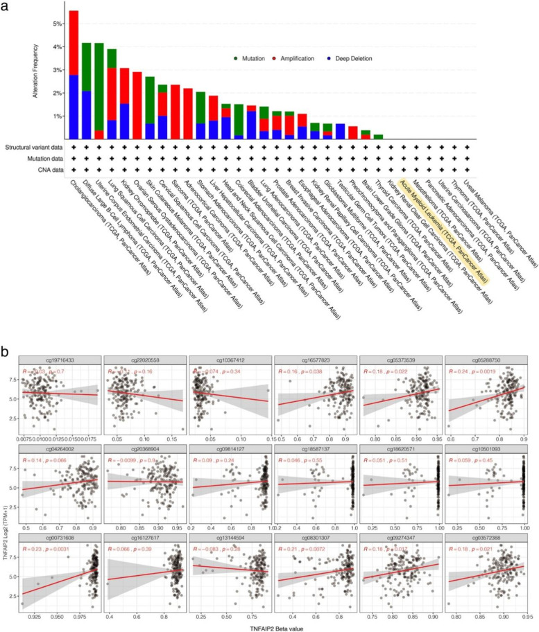
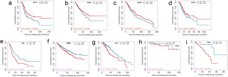
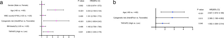
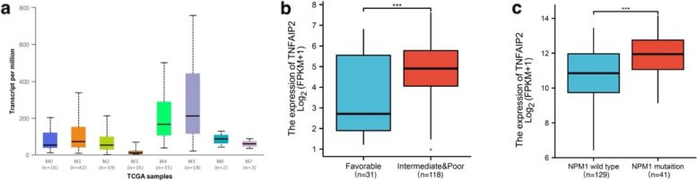
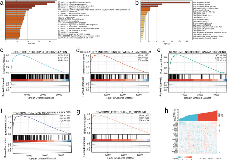
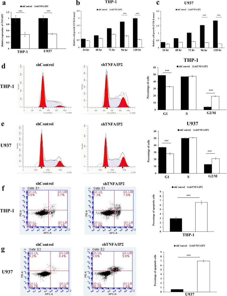
Results: By large-scale data mining, our study indicated that TNFAIP2 was differentially expressed across different normal and tumor tissues. TNFAIP2 expression was significantly increased in AML, particularly in French-American-British (FAB) classification M4/M5 patients, compared with corresponding control tissues. Overexpression of TNFAIP2 was an independent poor prognostic factor of overall survival (OS) and was associated with unfavorable cytogenetic risk and gene mutations in AML patients. DNA hypermethylation of TNFAIP2 at gene body linked to upregulation of TNFAIP2 and inferior OS in AML. Functional enrichment analysis indicated immunomodulation function and inflammation response of TNFAIP2 in leukemogenesis. Finally, the suppression of TNFAIP resulted in inhibition of proliferation by altering cell-cycle progression and increase of cell death by promoting early and late apoptosis in THP-1 and U937AML cells.

Conclusion: Collectively, the oncogenic TNFAIP2 can function as a novel biomarker and prognostic factor in AML patients. The immunoregulation function of TNFAIP2 warrants further validation in AML.

Keywords: Acute myeloid leukemia; Immunomodulation; Oncogenic; Prognosis; TNFAIP2.


Conflict of interest statement

The authors declare that they have no competing interests.

Figures

Fig. 1
Fig. 1
Differential expression of TNFAIP2 in cancer tissues and normal counterparts from TCGA and GTEx databases, analyzed by Mann–Whitney U test. *P <  0.05, **P < 0.01, ***P < 0.001. (LAML, acute myeloid leukemia; ACC, adrenalcortical carcinoma; BLCA, bladder urothelial carcinoma; BRCA, breast invasive carcinoma; CHOL, cholangiocarcinoma; COAD, colon adenocarcinoma; DLBC, diffuse large B-cell lymphoma; ESCA, esophageal carcinoma; GBM, glioblastoma multiforme; HNSC, Head and Neck squamous cell carcinoma; KICH, kidney chromophobe; KIRC, kidney renal clear cell carcinoma; KIRP, kidney renal papillary cell carcinoma; LGG, brain lower grade glioma; LIHC, liver hepatocellular carcinoma; LUAD, lung adenocarcinoma; LUSC, lung squamous cell carcinoma; MESO, mesothelioma; OV, ovarian serous cystadenocarcinoma; PAAD, pancreatic adenocarcinoma; PCPG, pheochromocytoma and paraganglioma; PRAD, prostate adenocarcinoma; READ, rectum adenocarcinoma; SARC, sarcoma; SKCM, skin cutaneous melanoma; STAD, stomach adenocarcinoma; TGCT, testicular germ cell tumor; THCA, thyroid carcinoma; THYM, thymoma; UCEC, uterine corpus endometrial carcinoma; UVM, uveal melanoma)
Fig. 2
Fig. 2
Genetic and epigenetic alterations of TNFAIP2 in AML patients from TCGA dataset. a Genetic features of TNFAIP2, including mutation, amplification, and deletion, in different tumors of TCGA analyzed by cBioPortal. b Correlation of TNFAIP2 methylation level and gene expression in AML patients analyzed by SMART App
Fig. 3
Fig. 3
Analysis of the prognostic value of TNFAIP2 expression on overall survival (OS) in different types of cancers by Timer 2.0 database. a BLAC, (b) SARC, (c) SKCM, (d) STAD, (e) LAML, (f) KIRC, (g) LGG, (h) THYM, (i) UVM. (LAML, acute myeloid leukemia; BLCA, bladder urothelial carcinoma; KIRP, kidney renal papillary cell carcinoma; LGG, brain lower grade glioma; SARC, sarcoma; SKCM, skin cutaneous melanoma; STAD, stomach adenocarcinoma; THYM, thymoma; UVM, uveal melanoma)
Fig. 4
Fig. 4
TNFAIP2 overexpression as an independent prognostic factor in AML patients. a Forest plot for univariate cox regression analysis of TNFAIP2 mRNA expression with OS in AML with different clinicopathological features. b Forest plot for multivariate cox regression analysis of TNFAIP2 mRNA expression with OS in AML with different clinicopathological features
Fig. 5
Fig. 5
TNFAIP2 expression and clinical features in AML patients in TCGA dataset. a Comparison of TNFAIP2 expression level among different subtypes of AML in the distribution of FAB classifications analyzed by UALCAN. b Comparison of TNFAIP2 expression level in AML patients according to cytogenetic risk stratification. c Comparison of TNFAIP2 expression level in AML patients according to NPM1 gene mutation status
Fig. 6
Fig. 6
Genome-wide genes associated with TNFAIP2 expression in AML. a Volcano plot for the co-expressed genes associated with TNFAIP2 expression, analyzed by LinkedOmics. b Volcano plot for differentially expressed genes (DEGs) between TNFAIP2high and TNFAIP2low groups. c Venn diagram for the overlapping genes between significantly co-expressed genes and significantly DEGs of TNFAIP2
Fig. 7
Fig. 7
Functional enrichment analysis of overlapping genes in AML. a Enrichment analysis of GO and KEGG pathway associated with TNFAIP2 expression analyzed by Metascape; b Enrichment analysis of overlapping genes in tissues and cells analyzed by Metascape; c-g Signaling pathways enriched by GSEA analyses of overlapping genes. h Co-expression of TNFAIP2 and immune-related genes in AML
Fig. 8
Fig. 8
Protein-protein interaction (PPI) network of overlapping genes in AML. a PPI network for the overlapping genes, analyzed by String; b Network of hub genes screened from overlapping genes by MCODE analysis with Cytoscape software
Fig. 9
Fig. 9
Oncogenic function of TNFAIP2 in AML cells. a qRT-PCR was performed to examine the efficiency of TNFAIP2 silencing in short hairpin RNA-stably transduced AML cell lines THP-1 and U937 at 120-hour post-transfection. b, c Cellular proliferation upon silencing of TNFAIP2 in THP-1 (b) and U937 (c) cells was studied by CCK-8 assay after transfection. d, e The cell-cycle distribution upon silencing of TNFAIP2 in THP-1 (d) and U937 (e) cells was studied by flow cytometry at 120-hour post-transfection. f, g The cell apoptosis upon silencing of TNFAIP2 in THP-1 (f) and U937 (g) cells was studied by flow cytometry at 120-hour post-transfection. Columns represented mean +/− 1SD from three independent experiments

References

    1. Juliusson G. Older patients with acute myeloid leukemia benefit from intensive chemotherapy: an update from the Swedish acute leukemia registry. Clin Lymphoma Myeloma Leuk. 2011;11(Suppl 1):S9–54. - PubMed
    1. Döhner H, Estey E, Grimwade D, Amadori S, Appelbaum FR, Büchner T, et al. Diagnosis and management of AML in adults: 2017 ELN recommendations from an international expert panel. Blood. 2017;129:424–447. doi: 10.1182/blood-2016-08-733196. - DOI - PMC - PubMed
    1. Tabata R, Chi S, Yuda J, Minami Y. Emerging immunotherapy for acute myeloid leukemia. Int J Mol Sci. 2021;22(4):1944. doi: 10.3390/ijms22041944. - DOI - PMC - PubMed
    1. Yang H, Bueso-Ramos C, DiNardo C, Estecio MR, Davanlou M, Geng QR, et al. Expression of PD-L1, PD-L2, PD-1 and CTLA4 in myelodysplastic syndromes is enhanced by treatment with hypomethylating agents. Leukemia. 2014;28:1280–1288. doi: 10.1038/leu.2013.355. - DOI - PMC - PubMed
    1. Zhou Q, Munger ME, Veenstra RG, Weigel BJ, Hirashima M, Munn DH, et al. Coexpression of Tim-3 and PD-1 identifies a CD8+ T-cell exhaustion phenotype in mice with disseminated acute myelogenous leukemia. Blood. 2011;117:4501–4510. doi: 10.1182/blood-2010-10-310425. - DOI - PMC - PubMed