

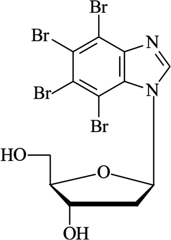
Antitumor activity of the protein kinase inhibitor 1-(β-D-2'-deoxyribofuranosyl)-4,5,6,7-tetrabromo- 1H-benzimidazole in breast cancer cell lines
- PMID: 36243702
- PMCID: PMC9571492
- DOI: 10.1186/s12885-022-10156-8
Antitumor activity of the protein kinase inhibitor 1-(β-D-2'-deoxyribofuranosyl)-4,5,6,7-tetrabromo- 1H-benzimidazole in breast cancer cell lines
Abstract
Background: The protein kinases CK2 and PIM-1 are involved in cell proliferation and survival, the cell cycle, and drug resistance, and they are found overexpressed in virtually all types of human cancer, including breast cancer. In this study, we investigated the antitumor activity of a deoxynucleoside derivative, the protein kinase inhibitor compound 1-(β-D-2'-deoxyribofuranosyl)-4,5,6,7-tetrabromo-1H-benzimidazole (K164, also termed TDB), inter alia CK2 and PIM-1, on breast cancer cell lines (MDA-MB-231, MCF-7, and SK-BR-3).
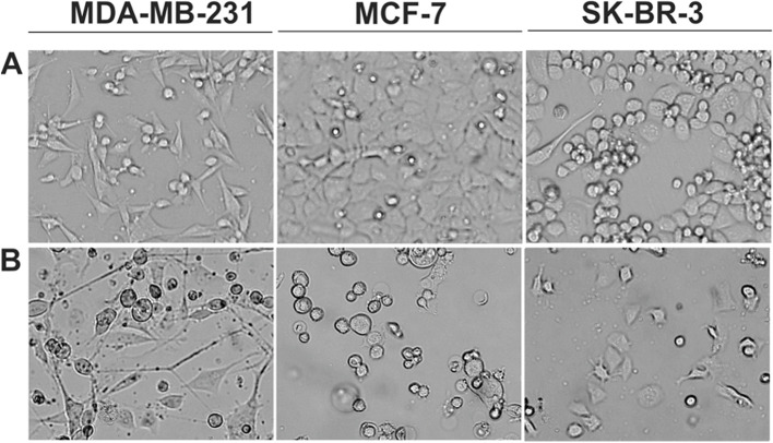
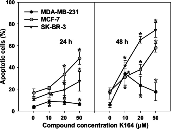
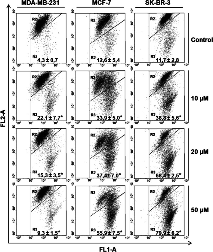
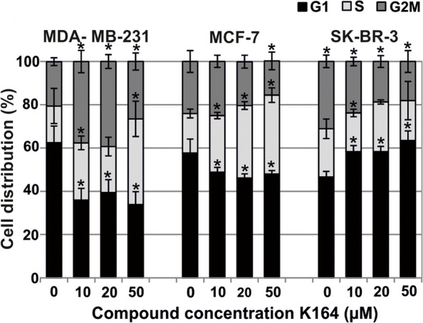
Methods: An evaluation of the cytotoxic and proapoptotic effects, mitochondrial membrane potential (ΔΨm), and cell cycle progression was performed using an MTT assay, flow cytometry, and microscopic analysis. The Western blotting method was used to analyze the level of proteins important for the survival of breast cancer cells and proteins phosphorylated by the CK2 and PIM-1 kinases.
Results: The examined compound demonstrated the inhibition of cell viability in all the tested cell lines and apoptotic activity, especially in the MCF-7 and SK-BR-3 cells. Changes in the mitochondrial membrane potential (ΔΨm), cell cycle progression, and the level of the proteins studied were also observed.
Conclusions: The investigated CK2 and PIM-1 kinase inhibitor K164 is a promising compound that can be considered a potential agent in targeted therapy in selected types of breast cancer; therefore, further research is necessary.
Keywords: 1-(β-D-2′-deoxyribofuranosyl)-4,5,6,7-tetrabromo-1H-benzimidazole; Apoptosis; Breast cancer cell lines; Flow cytometry; Protein kinase inhibitor.
© 2022. The Author(s).
Conflict of interest statement
The authors declare that they have no competing interests.
Figures


References
-
- Tawfic S, Yu S, Wang H, Faust R, Davis A, Ahmed K. Protein kinase CK2 signal in neoplasia. Histol Histopathol. 2001;16:573–582. - PubMed
MeSH terms
Substances
LinkOut - more resources
Full Text Sources
Medical
Miscellaneous
