. 2022 Oct 15;17(1):453.
doi: 10.1186/s13018-022-03334-8.

miR-124-3p sabotages lncRNA MALAT1 stability to repress chondrocyte pyroptosis and relieve cartilage injury in osteoarthritis

Affiliations

miR-124-3p sabotages lncRNA MALAT1 stability to repress chondrocyte pyroptosis and relieve cartilage injury in osteoarthritis

Rigbat Rozi et al. J Orthop Surg Res. .

Abstract

Background: Osteoarthritis (OA) is a prevalent inflammatory joint disorder. microRNAs (miRNAs) are increasingly involved in OA.

Aim: Our study is proposed to clarify the role of miR-124-3p in chondrocyte pyroptosis and cartilage injury in OA.

Methods: OA mouse model was established via the treatment of destabilization of the medial meniscus (DMM), and the in vitro cell model was also established as mouse chondrocytes were induced by lipopolysaccharide (LPS). Mouse cartilage injury was assessed using safranin-O-fast green staining, hematoxylin-eosin staining, and OARSI grading method. Expressions of miR-124-3p, MALAT1, KLF5, and CXCL11 were determined. Cartilage injury (MMP-13, osteocalcin), inflammation (IL-6, IL-2, TNF-, IL-1β, and IL-18)- and pyroptosis-related factors (Cleaved Caspase-1 and GSDMD-N) levels were detected. Mechanically, MALAT1 subcellular localization was confirmed. The binding relationships of miR-124-3p and MALAT1 and MALAT1 and KLF5 were verified. MALAT1 half-life period was detected. Then, miR-124-3p was overexpressed using agomiR-124-3p to perform the rescue experiments with oe-MALAT1 or oe-CXCL11.

Results: miR-124-3p was downregulated in DMM mice and LPS-induced chondrocytes where cartilage injury, and increased levels of inflammation- and pyroptosis-related factors were found. miR-124-3p overexpression relieved cartilage injury and repressed chondrocyte pyroptosis. miR-124-3p bounds to MALAT1 to downregulate its stability and expression, and MALAT1 bounds to KLF5 to enhance CXCL11 transcription. Overexpression of MALAT1 or CXCL11 annulled the repressive function of miR-124-3p in chondrocyte pyroptosis.

Conclusion: miR-124-3p reduced MALAT1 stability and inhibited the binding of MALAT1 and KLF5 to downregulate CXCL11, thereby suppressing chondrocyte pyroptosis and cartilage injury in OA.

Keywords: Chondrocytes; LncRNA MALAT1; LncRNA stability; Osteoarthritis; Pyroptosis; microRNA-124-3p.


Conflict of interest statement

The authors report there are no competing interests to declare.

Figures
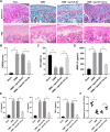
Fig. 1
Fig. 1
miR-124-3p overexpression alleviates OA mouse cartilage injury. DMM mouse model was established to mimic OA mice, which were injected with agomiR-124-3p to overexpress miR-124-3p, with agomiR-NC as the control. A, mouse knee cartilage damage was determined by Safranin-O-fast green staining and H&E staining. B, degree of mouse knee cartilage damage was evaluated by OARSI grading. C, D, and E, contents of OC, MMP-13, IL-6, IL-2, and TNF-α in mouse serum were detected by ELISA. F, miR-124-3p expression in tissue was measured by RT-qPCR. N = 6. The data in panels BE were presented as mean ± standard deviation. One-way ANOVA was used to analyze the data in panels B, C, D, E, and F. Tukey's multiple comparisons test was applied for the post hoc test. *p < 0.05, **p < 0.01
Fig. 2
Fig. 2
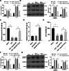
miR-124-3p overexpression represses mouse chondrocyte pyroptosis in vitro and in vivo. agomiR-124-3p was injected into OA mouse knee or transfected into mouse chondrocytes, with agomiR-NC as the control. A, expressions of IL‐1β and IL‐18 in the serum were detected by ELISA. B, expressions of Cleaved Caspase-1 and GSDMD-N in mouse tissue were detected by western blot analysis. Inflammatory injury in mouse chondrocytes was induced by LPS. C and D, miR-124-3p expression in chondrocytes (C) and miR-124-3p transfection efficiency (D) were measured by RT-qPCR. E, cell activity was detected by CCK-8 method. F, expressions of IL‐1β and IL‐18 in cells were detected by ELISA. G, expressions of Cleaved Caspase-1 and GSDMD-N in cells were detected by western blot analysis. N = 6, cell experiments were repeated 3 times independently. The results were presented as mean ± standard deviation. Two-way ANOVA was used to analyze the data in panels A, B, F, and G, and one-way ANOVA was used to analyze the data in panels C, D, and E. Tukey's multiple comparisons test was applied for the post hoc test. **p < 0.01
Fig. 3
Fig. 3
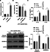
miR-124-3p directly binds to MALAT1 to reduce its stability and expression in OA. A, the binding relation between miR-124-3p and MALAT1 was revealed through the StarBase website. B and C, the binding relation between miR-124-3p and MALAT1 was verified by dual-luciferase reporter gene assay (B) and RIP assay (C). D, half-life period of MALAT1 (as presented by the dotted line) in cells with miR-124-3p overexpression after the treatment of actinomycin D. E and F, MALAT1 expression in tissue (E) and chondrocytes (F). N = 6, cell experiments were repeated 3 times independently. The data in panels BD, and F were presented as mean ± standard deviation. One-way ANOVA was used to analyze the data in panels E and F, and two-way ANOVA was used to analyze the data in panels B, C, and D, and one. Tukey's multiple comparisons test was applied for the post hoc test. *p < 0.05, **p < 0.01
Fig. 4
Fig. 4
MALAT1 overexpression reverses the inhibiting role of miR-124-3p overexpression in chondrocyte pyroptosis. oe-MALAT1 was transfected into cells to upregulate MALAT1 expression to conduct combined experiments with agomiR-124-3p, with oe-NC transfection as the control. A, MALAT1 transfection efficiency was verified by RT-qPCR. B, cell activity was detected by CCK-8 method. C, expressions of IL‐1β and IL‐18 in mouse cartilage tissue were detected by ELISA. D, expressions of Cleaved Caspase-1 and GSDMD-N in mouse cartilage tissue were detected by western blot analysis. Cell experiments were repeated 3 times independently. The results were presented as mean ± standard deviation. One-way ANOVA was used to analyze the data in panels A and B, and two-way ANOVA was used to analyze the data in panels C and D. Tukey's multiple comparisons test was applied for the post hoc test. **p < 0.01
Fig. 5
Fig. 5
MALAT1 binds to transcription factor KLF5 to promote CXCL11 transcription. A, the subcellular localization of MALAT1 was predicted via the LncATLAS website. B, fractionation of nuclear and cytoplasmic RNA verified that MALAT1 was mainly localized in the nucleus. C, The binding relations between MALAT1 and KLF5 and between KLF5 and CXCL11 were predicted through the RNAInter database. D and E, the binding relation between KLF5 and MALAT1 was verified by RIP assay (D) and RNA pull-down assay (E). si-KLF5-1 or si-KLF5-2 was transfected into cells to downregulate KLF5 expression, with si-NC as the control. F, KLF5 transfection efficiency was verified by RT-qPCR. G, CXCL11 expression in cells with si-KLF5 was detected by RT-qPCR. H and I, KLF5 expression in tissue (H) and chondrocytes (I) were determined by RT-qPCR. J and K, CXCL11 expression in tissue (J) and chondrocytes (K) were determined by RT-qPCR N = 6, cell experiments were repeated 3 times independently. Data in panels BD, EG, I, and K were presented as mean ± standard deviation. Two-way ANOVA was used to analyze the data in panel D, and one-way ANOVA was used to analyze the data in panels EK. Tukey's multiple comparisons test was applied for the post hoc test. *p < 0.05, **p < 0.01
Fig. 6
Fig. 6
CXCL11 overexpression reverses the inhibiting role of miR-124-3p overexpression in chondrocyte pyroptosis. oe-CXCL11 was transfected into cells to upregulate CXCL11 expression to conduct combined experiments with agomiR-124-3p. A, CXCL11 transfection efficiency was verified by RT-qPCR. B, cell activity was detected by CCK-8 method. C, expressions of IL‐1β and IL‐18 in mouse cartilage tissue were detected by ELISA. D, expressions of Cleaved Caspase-1 and GSDMD-N in mouse cartilage tissue were detected by western blot analysis. Cell experiments were repeated 3 times independently. The results were presented as mean ± standard deviation. One-way ANOVA was used to analyze the data in panels A and B, and two-way ANOVA was used to analyze the data in panels C and D. Tukey's multiple comparisons test was applied for the post hoc test. *< 0.05, **p < 0.01
Fig. 7
Fig. 7
The mechanism of miR-124-3p in OA. miR-124-3p bound to MALAT1 to reduce its stability and expression, repressed the binding of MALAT1 and the transcription factor KLF5, and suppressed CXCL11 transcription, thereby inhibiting chondrocyte pyroptosis and cartilage damage in OA

Similar articles

Cited by

References

    1. Bijlsma JW, Berenbaum F, Lafeber FP. Osteoarthritis: an update with relevance for clinical practice. Lancet. 2011;377(9783):2115–2126. doi: 10.1016/S0140-6736(11)60243-2. - DOI - PubMed
    1. O'Neill TW, McCabe PS, McBeth J. Update on the epidemiology, risk factors and disease outcomes of osteoarthritis. Best Pract Res Clin Rheumatol. 2018;32(2):312–326. doi: 10.1016/j.berh.2018.10.007. - DOI - PubMed
    1. Glyn-Jones S, Palmer AJ, Agricola R, Price AJ, Vincent TL, Weinans H, et al. Osteoarthritis Lancet. 2015;386(9991):376–387. - PubMed
    1. Sacitharan PK. Ageing and osteoarthritis. Subcell Biochem. 2019;91:123–159. doi: 10.1007/978-981-13-3681-2_6. - DOI - PubMed
    1. Shen J, Abu-Amer Y, O'Keefe RJ, McAlinden A. Inflammation and epigenetic regulation in osteoarthritis. Connect Tissue Res. 2017;58(1):49–63. doi: 10.1080/03008207.2016.1208655. - DOI - PMC - PubMed

MeSH terms