. 2023 Jan:359:114252.
doi: 10.1016/j.expneurol.2022.114252. Epub 2022 Oct 13.

Traumatic axonopathy in spinal tracts after impact acceleration head injury: Ultrastructural observations and evidence of SARM1-dependent axonal degeneration

Affiliations

Traumatic axonopathy in spinal tracts after impact acceleration head injury: Ultrastructural observations and evidence of SARM1-dependent axonal degeneration

Athanasios S Alexandris et al. Exp Neurol. 2023 Jan.

Abstract

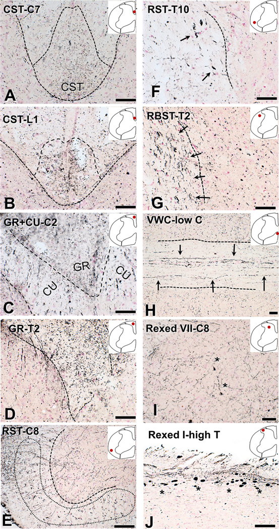
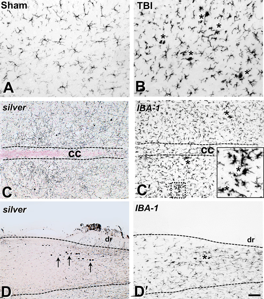
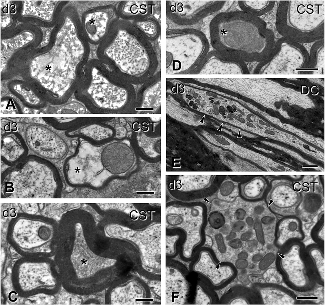
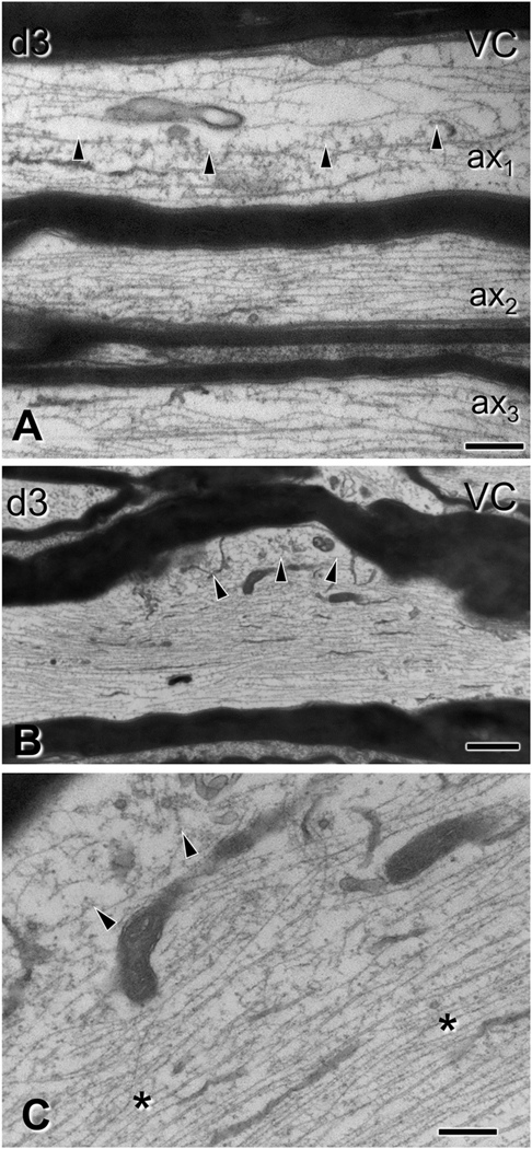
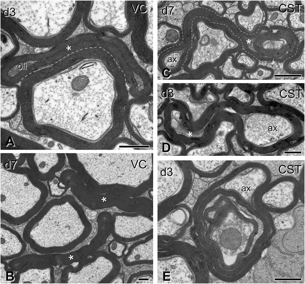
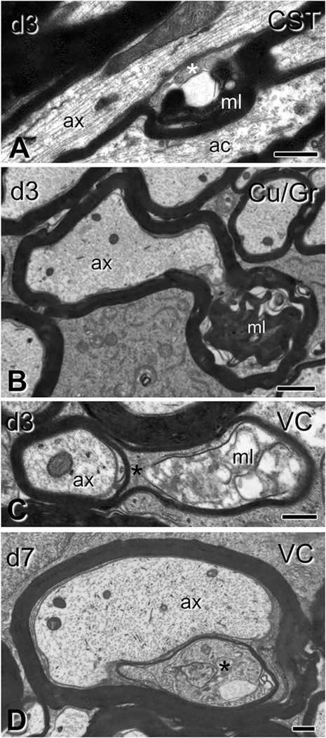
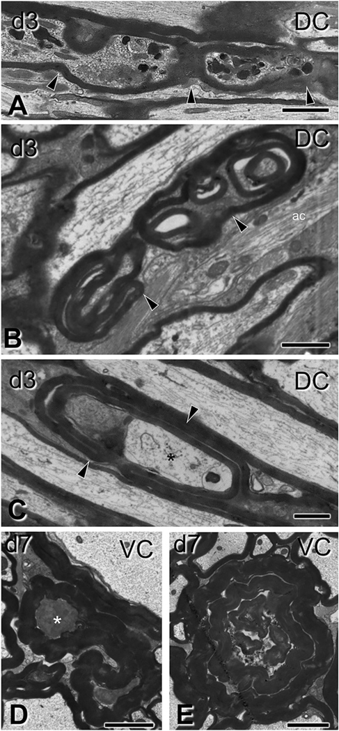
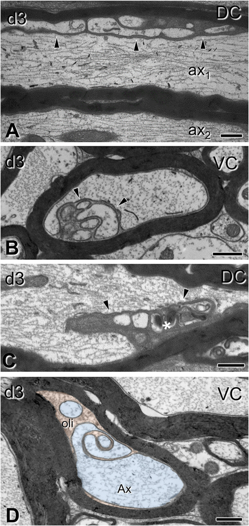
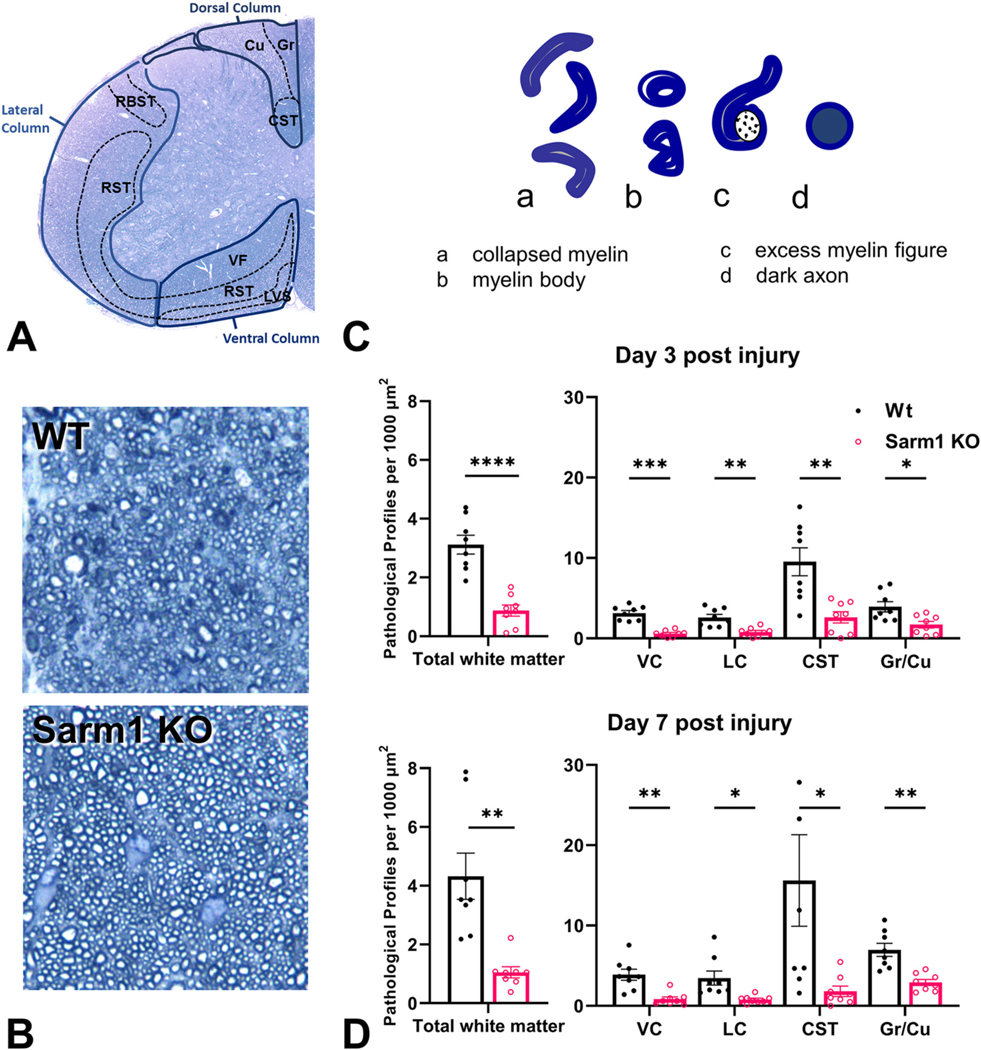
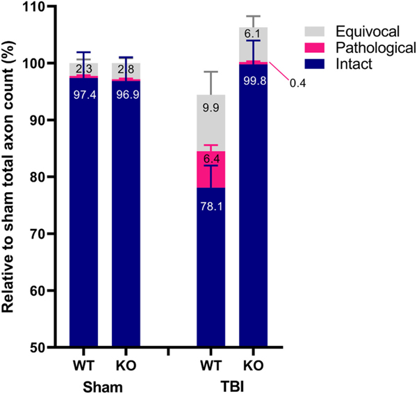
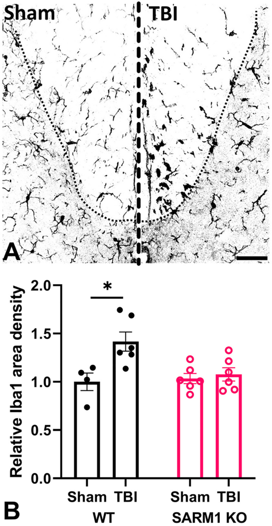
Traumatic axonal injury (TAI) and the associated axonopathy are common consequences of traumatic brain injury (TBI) and contribute to significant neurological morbidity. It has been previously suggested that TAI activates a highly conserved program of axonal self-destruction known as Wallerian degeneration (WD). In the present study, we utilize our well-established impact acceleration model of TBI (IA-TBI) to characterize the pathology of injured myelinated axons in the white matter tracks traversing the ventral, lateral, and dorsal spinal columns in the mouse and assess the effect of Sterile Alpha and TIR Motif Containing 1 (Sarm1) gene knockout on acute and subacute axonal degeneration and myelin pathology. In silver-stained preparations, we found that IA-TBI results in white matter pathology as well as terminal field degeneration across the rostrocaudal axis of the spinal cord. At the ultrastructural level, we found that traumatic axonopathy is associated with diverse types of axonal and myelin pathology, ranging from focal axoskeletal perturbations and focal disruption of the myelin sheath to axonal fragmentation. Several morphological features such as neurofilament compaction, accumulation of organelles and inclusions, axoskeletal flocculation, myelin degeneration and formation of ovoids are similar to profiles encountered in classical examples of WD. Other profiles such as excess myelin figures and inner tongue evaginations are more typical of chronic neuropathies. Stereological analysis of pathological axonal and myelin profiles in the ventral, lateral, and dorsal columns of the lower cervical cord (C6) segments from wild type and Sarm1 KO mice at 3 and 7 days post IA-TBI (n = 32) revealed an up to 90% reduction in the density of pathological profiles in Sarm1 KO mice after IA-TBI. Protection was evident across all white matter tracts assessed, but showed some variability. Finally, Sarm1 deletion ameliorated the activation of microglia associated with TAI. Our findings demonstrate the presence of severe traumatic axonopathy in multiple ascending and descending long tracts after IA-TBI with features consistent with some chronic axonopathies and models of WD and the across-tract protective effect of Sarm1 deletion.

Keywords: Corticospinal tract; Electron microscopy; Microglia; Myelin; Neurodegeneration; Neuropathy; Oligodendrocyte; Traumatic axonal injury; Wallerian degeneration.


Conflict of interest statement

Declaration of Competing Interest None.

Figures

Fig. 1.
Fig. 1.
Degeneration of ventral, lateral and dorsal spinal columns after impact acceleration injury revealed with amino-cupric silver at 3 days. With the exception of (E), dotted lines mark white-gray matter border or midline. The anatomical location is indicated in each panel with an inset and spinal segments are denoted with letters and numbers (C, cervical; T, thoracic; L, lumbar). Approximate tract delineation is based on the map of Watson and Harrison (2012). (A-B) Degenerative changes in the corticospinal tract in coronal sections. (C–D). Degenerating fibers in the gracilis and cuneatus tracts in coronal. (E-F) Degenerative changes in large-caliber axons in the ventral and lateral columns roughly corresponding to the reticulospinal tract (arrows). (G) Coronal section through the thoracic cord showing silver precipitation in large fibers corresponding to the rubrospinal tract (arrows). (H) Horizontal section through the ventral white commissure in the lower cervical cord showing large degenerating fibers putatively belonging to the reticulospinal tract (arrows). (I) Fine silver precipitation in neuropil in Rexed VII of a coronal section through the lower cervical cord. (J) Argyrophilic axonal spheroids in the dorsal root entry zone (Rexed I) in a horizontal section through higher thoracic cord. Asterisks indicate pathological axon swellings and bulbs. CST, corticospinal tract; CU, cuneatus; GR, gracilis; RBST, rubrospinal tract; RST, reticulospinal tract; VWC, ventral white commissure. Scale bars: 50 μm.
Fig. 2.
Fig. 2.
Traumatic brain injury is associated with microglial activation in the spinal cord tracts. (A-B) IBA1 immunohistochemistry labels resting microglia in the mid-cervical ventral column of sham-injured mice (A) and the deramified microglia and microglial nodules (asterisks) 3 days after IA-TBI (B). (C-C) Adjacent horizontal sections through Rexed VII at a mid-cervical level stained for cupric silver (C) and IBA1 immunoreactivity (C). Note the widespread fine neuropil silver precipitation in terminals in (C) and extensive microglial activation with the presence of microglial nodules (asterisks and inset) in (C). (D-D) Silver (D) and IBA1 (D) staining of adjacent horizontal sections through Rexed I at an upper thoracic level Arrows in panel (D) indicate the presence of large axonal spheroids in the root entry zone; same area is featured by deramified IBA1(+) profiles in panel (D). cc, central canal; dr, dorsal root. Scale bars: 100 μm.
Fig. 3.
Fig. 3.
Gross alterations of spinal axons 3 days after impact acceleration injury. Electron micrographs are from sections through the corticospinal tract (A-D, F) or the dorsal columns (E) corresponding to C6–7 segments. (A-B) Transverse axonal profiles showing hydropic changes (asterisks) with dissolution of structure at different degrees, and the presence of distended mitochondria (B). Note the thin myelin sheath in the pathological axon in (B). (C–D) Transversely sectioned profiles with axoplasmic flocculation (C) and advanced condensation (D; asterisks). In both cases, myelin sheath appears normal. (E-F) Accumulation of mitochondria, inclusions and dense bodies in distended segments of longitudinally (E) and transversely (F) sectioned axons (arrowheads). Note the relatively thin myelin sheath in (E) and the absence of myelin sheath in (F). CST, corticospinal tract; DC, dorsal column. Scale bars: A-C, 400 nm; D-E, 500 nm; F, 1 μm.
Fig. 4.
Fig. 4.
Subtle alterations of spinal cord axons 3 days after impact acceleration injury. Electron micrographs are from sections through the ventral column corresponding to C6–7 segments. (A-B) Regional rarefaction or deformation of microtubule content in ax1 (A, arrowheads; also throughout ax3) and in an axonal protrusion/varicosity of the axon illustrated in (B, arrowheads). Compare with normal axons (e.g. ax2 in A). The myelin sheath of pathological axons is often compact, indistinguishable from that in non-injured axons. (C) Higher magnification of (B), showing localized disruption of the cytoskeleton (arrowheads) and compaction of neurofilaments in the underlying region (asterisks). VC, ventral column. Scale bars; A, C, 400 nm; B, 1 μm.
Fig. 5.
Fig. 5.
Gross myelin pathology in the form of collapsed myelin or excess myelin profiles. Electron micrographs are from sections through the ventral column in panels (A-B) and the corticospinal tract in panels (C-E), (C6–7 level). (A-B) Collapsed myelin profiles, i.e., myelin sheaths closely apposed to each other with little or no tissue between them, are seen at 3 days (A) and, more commonly, at 7 days (B) post-injury (asterisks). Oli, oligodendrocyte. (C-E) Excess myelin profiles appear as morphologically complex tube-like structures with morphological similarities to collapsed myelin objects. These profiles loop around normal-looking or abnormal axons or oligodendrocyte cytoplasm. Note the ensheathing of normal axonal material (ax) in (C), but also surrounding of tissue with electron density typical of oligodendrocytes (C–D, asterisks). These various tissues may be contained in the same enclosure (C,D). Excess myelin often wraps itself in concentric loops containing normal and abnormal tissue elements in the form of tomacula (E). CST, corticospinal tract; VC, ventral column. Scale bars: A, B, D, E, 500 nm; C, 800 nm.
Fig. 6.
Fig. 6.
Subtle myelin pathology after traumatic brain injury. Electron micrographs are from sections through the corticospinal tract (A), gracilis/cuneatus (B) and ventral column (C–D) (C6–7 level). (A-B) In longitudinal (A) and transverse (B) sections, loose and more compact myelin loops (ml) form ectopically between intact axons (ax) and apparently intact sheaths, with/without oligodendrocyte cytoplasm (asterisk in A). (C–D) Here the myelin sheath of normal-looking axons is split by a massive accumulation of dense oligodendrocyte cytoplasm (asterisks). In (C) there is also disorderly loose myelin (ml). ac, astrocyte; CST, corticospinal tract; Gr/Cu, gracilis and cuneatus; VC, ventral column. Scale bars: A, 600 nm; B, 1 μm; C, 250 nm; D-E, 500 nm.
Fig. 7.
Fig. 7.
Ovoid-like formation and axonal fragmentation after traumatic brain injury. Electron micrographs are from sections through the dorsal columns (A-C), and ventral columns (D-E) (C6–7 level). (A) A longitudinally cut axon showing accumulation of mitochondria and dense bodies (see also Fig. 3 E–F) but also periodic segmental constriction of the axon (arrowheads), typical of early Wallerian degeneration in peripheral axons. (B–C) Ovoid formations in longitudinal sections of myelinated axons (arrows) with (C) or without (B) the preservation of portions of hydropic axoplasm (asterisk). In both panels, note the complex arrangement of compact-appearing myelin sheaths. (D-E) Profiles of myelin bodies that may represent a later stage of degeneration at 7 days post- injury. There is loss of periodicity in myelin sheaths. ac, astrocyte; DC, dorsal column; VC, ventral column. Scale bars: A, D, E, 2 μm; B–C, 1 μm.
Fig. 8.
Fig. 8.
Oligodendrocyte cytoplasm protrusions into the main axon space seen after traumatic brain injury. Electron micrographs are from sections through the dorsal columns (A,C) and ventral columns (B, D) (C6–7 level). (A) Lobular evaginations of oligodendrocyte cytoplasm. These entities are separated from axons via intact membranes (arrowheads). Some axons have cytoskeletal disorganization (ax1 in A;); compare with a normal axon without evident affiliation with such evaginations (ax2 in A). (B) Cross-sectional configuration of evaginations similar to the ones in (A). (C) Some evaginations contain dark oligodendrocyte cytoplasm (arrowhead) and vacuolar or hyperdense inclusions (asterisk). (D) Axon showing evidence of evagination process. The inner tongue of the oligodendrocyte (oli; shown in orange), intends deeply the axon and partially partitions the axoplasm (Ax; light blue). Note the degenerative flocculation and absence of microtubules in the axoplasm. DC, dorsal column; VC, ventral column. Scale bars: 500 nm. (For interpretation of the references to colour in this figure legend, the reader is referred to the web version of this article.)
Fig. 9.
Fig. 9.
The effects of Sarm1 knockout on pathological axonal degeneration in the spinal cord after impact acceleration traumatic brain injury. (A) Toluidine blue-stained semithin transverse section of mouse cervical spinal cord with annotations of white matter tracts based on Watson and Harrison (2012). White matter tracts counted in this study are delineated with solid blue lines (DC, LC, VC). (B) Representative images from wt and Sarm1 KO mice through the CST 3 days after injury. (C) Sketches of main types of pathological profiles unequivocally visualized on semithin material in this study. (D) Pathological profile densities (number of pathological profiles per mm2) in the lower cervical spinal cord (C6–7) 3 and 7 days after IA-TBI, in WT and SARM1 KO mice. *p < 0.05, **p < 0.01, ***p < 0.001, ****p < 0.0001. CST, corticospinal tract; Cu, Cuneatus; Gr, gracilis; LC, lateral column; lvs, lateral vestibulospinal tract; rbst, rubrospinal tract; rst-c, caudal reticulospinal tract; rst-v, ventral reticulospinal tract; VC, ventral column; vf, ventral funiculus. (For interpretation of the references to colour in this figure legend, the reader is referred to the web version of this article.)
Fig. 10.
Fig. 10.
Stereological analysis of apparently intact, pathological and equivocal axonal profiles in the corticospinal tract of wt and Sarm1 KO mice 7 days after injury. Values are expressed relative to the total axon counts of sham animals for each genotype. Sarm1 knockout leads to significant protection of intact axons (t13.9 = 3.8 p = 0.002) and significant reduction in pathological profiles (t8 = 4.39; p = 0.002) compared to injured wt mice.
Fig. 11.
Fig. 11.
Sarm1 knockout ameliorates TBI-induced microglial reactivity in the corticospinal tract. (A) Representative confocal images of IBA1 immunoreactivity in the CST of wt animals 7 days after TBI and sham injury. (B) Changes in the areal density of IBA1 immunoreactivity in the corticospinal tract of wt and Sarm1 KO mice after TBI compared to sham-injured mice for each genotype. In wt, but not Sarm1 KO mice, there is significant increase in IBA1 immunoreactivity after TBI, t8 = 2.90, p = 0.018 (*).

Similar articles

Cited by

References

    1. Adams JH, Doyle D, Graham DI, Lawrence AE, McLellan DR, 1984. Diffuse axonal injury in head injuries caused by a fall. Lancet 2 (8417–8418), 1420–1422. - PubMed
    1. Alexandris AS, Ryu J, Rajbhandari L, Harlan R, McKenney J, Wang Y, Aja S, Graham D, Venkatesan A, Koliatsos VE, 2022a. Protective effects of NAMPT or MAPK inhibitors and NaR on Wallerian degeneration of mammalian axons. Neurobiol. Dis. 171, 105808. - PMC - PubMed
    1. Alexandris AS, Wang Y, Frangakis CE, Lee Y, Ryu J, Alam Z, Koliatsos VE, 2022b. Long-term changes in axon calibers after injury: observations on the mouse corticospinal tract. Int. J. Mol. Sci. 23 (13). - PMC - PubMed
    1. Armstrong RC, Mierzwa AJ, Marion CM, Sullivan GM, 2016. White matter involvement after TBI: clues to axon and myelin repair capacity. Exp. Neurol. 275 (Pt 3), 328–333. - PubMed
    1. Bando Y, Nomura T, Bochimoto H, Murakami K, Tanaka T, Watanabe T, Yoshida S, 2015. Abnormal morphology of myelin and axon pathology in murine models of multiple sclerosis. Neurochem. Int. 81, 16–27. - PubMed

Publication types