. 2022 Jun 24:2022:7104592.
doi: 10.1155/2022/7104592. eCollection 2022.

APY0201 Represses Tumor Growth through Inhibiting Autophagy in Gastric Cancer Cells

Affiliations

APY0201 Represses Tumor Growth through Inhibiting Autophagy in Gastric Cancer Cells

Huan Li et al. J Oncol. .

Abstract

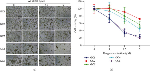
Gastric cancer (GC) is one of the most common cancers globally. There are currently few effective chemotherapeutic drugs available for GC patients. The inhibitors of phosphatidylinositol kinase containing an FYVE finger structure (PIKfyve) have shown significant anticancer effects in several types of cancers, but their effectiveness in GC remains unknown. In this study, we investigate the effect of APY0201, an inhibitor of PIKfyve, on GC tumor growth and its mechanism of action. It was found that APY0201 inhibited GC cell proliferation in in vitro GC cell model, organoid model, and in vivo xenograft tumor model. Through analyzing cell autophagy, we found that APY0201 might block autophagic flux by impairing lysosome degradation function of GC cells, inducing the accumulation of autophagosomes, thus causing the inhibition of GC cell proliferation. We also found that APY0201 induced G1/S phase arrest in GC cells. Importantly, APY0201 was also effective in inducing repression of autophagy and cell cycle arrest in the mouse tumor xenograft. Our results suggest that APY0201 could be a new promising therapeutic option for GC.


Conflict of interest statement

The authors declare no conflicts of interest.

Figures

Figure 1
Figure 1
APY0201 inhibits proliferation of GC cells. (a) Molecular structure of APY0201. (b) AGS, SGC7901, BGC823, MKN28, and SNU719 cells were treated with varied concentrations of APY0201 for 48 h and cell viability was determined. (c) Colony formation in AGS and SGC7901 cells treated with various concentrations of APY0201 for 14 days. (d)-(e) Statistical analysis of the numbers of colonies in part c. (f), (h) The EdU proliferation test was used to investigate the effects of varied concentrations of APY0201 on AGS and SGC7901 cells for 48 h. The scale bar is 300 μm. (g), (i) Proportions of EdU-positive cells in AGS and SGC7901 cells. P < 0.05, ∗∗P < 0.01, ∗∗∗P < 0.001, ∗∗∗∗P < 0.0001 compared with 0 μM APY0201 group.
Figure 2
Figure 2
APY0201 inhibits the growth of GC organoids. GC organoids were treated with various concentrations of APY0201 for 3 days. (a) Light microscope photos of GC organoids. The scale bar is 300 μm. (b) GC organoids were tested for cell viability by the CCK8 assay, and a broken line graph was created to illustrate this.
Figure 3
Figure 3
APY0201 inhibits the growth of GC xenografts. (a) Photographs of subcutaneous transplanted tumors and nude mice. (b) The average weight of transplanted tumors. (c) The volume of the transplanted tumor in each nude mouse was measured every 2 days, and a tumor growth curve during APY0201 treatment was plotted. (d) Mice were weighed every 2 days and a body weight change curve during APY0201 treatment was plotted.
Figure 4
Figure 4
APY0201 induces autophagosome accumulation. (a)-(b) AGS and SGC7901 cells were treated with APY0201 at various concentrations for 24 h and the expressions of LC3 and p62 were determined by western blotting. (c)-(d) AGS and SGC7901 cells were treated with DMSO and APY0201 with or without BafA1 for 24 h and the expressions of LC3 and p62 were determined by western blotting. (e), (g) AGS and SGC7901 cells stably expressing StubRFP-SensGFP-LC3 were seeded onto confocal culture dishes and treated with different drugs for 24 h. Confocal microscopy revealed the development of LC3 dots in AGS and SGC7901 cells (λex/green = 488 nm, λem/green = 500–530 nm; λex/red = 545 nm, λem/red = 590–630 nm). The scale bar is 25 μm. (f), (h) Histograms were based on the numbers of yellow and red dots in different drug groups shown in parts (e) and (g). P < 0.05, ∗∗P < 0.01, ∗∗∗P < 0.001, ∗∗∗∗P < 0.0001 were compared with autophagosomes in the control group; #P < 0.05, ##P < 0.01, ###P < 0.001, ####P < 0.0001 were compared with autolysosomes in the control group.
Figure 5
Figure 5
APY0201 impairs the degradation function of lysosomes by inhibiting the activity of cathepsin in GC cells. (a)-(b) AGS and SGC7901 cells were treated with APY0201 at various concentrations for 24 h and the expressions of CTSD and CTSB were detected by western blotting.
Figure 6
Figure 6
APY0201-induced autophagosome accumulation inhibits proliferation of GC cells. AGS and SGC7901 cells were infected with lentiviruses carrying shNC and shATG5 genes. Subsequent experiments were carried out with AGS and SGC7901 cells stably expressing shNC and shATG5. (a, b) After 24 h of treatment with DMSO and APY0201, AGS and SGC7901 cells were examined by western blotting for ATG5 and LC3 expression. (c), (d) DMSO and APY0201 were applied to AGS and SGC7901 cells for 48 h and their viability was determined by CCK8 assay. Histograms were then drawn. ∗∗∗∗P < 0.0001; ns, no statistically significant difference compared with the shNC group.
Figure 7
Figure 7
APY0201 induces G1/S phase arrest of GC cells. (a), (c) After 24 h of DMSO and APY0201 treatment, the cell cycle of AGS and SGC7901 cells was examined by flow cytometry. (b), (d) Percentage distribution of cell cycle phases in the control group and APY0201 group. P < 0.05, ∗∗P < 0.01; ns, no statistically significant difference compared with control group. (e), (f) AGS and SGC7901 cells were treated with APY0201 at various concentrations for 24 h. The expression levels of CDK2, CDK4, CDK6, cyclin D1, cyclin E1, p21, and p27 were analyzed by western blotting.
Figure 8
Figure 8
APY0201 inhibits the proliferation of GC transplanted tumor cells by interrupting autophagic flux and cell cycle arrest. (a) The expression of LC3, p62, and p21 in transplanted tumors was analyzed by western blotting. (b) The expression of Ki67, LC3, p62, and p21 in transplanted tumors was detected by immunohistochemistry. The scale bar is 200 μm.

References

    1. Sung H., Ferlay J., Siegel R. L., et al. Global cancer statistics 2020: GLOBOCAN estimates of incidence and mortality worldwide for 36 cancers in 185 countries. CA: A Cancer Journal for Clinicians . 2021;71(3):209–249. doi: 10.3322/caac.21660. - DOI - PubMed
    1. Smyth E. C., Nilsson M., Grabsch H. I., van Grieken N. C., Lordick F. Gastric cancer. The Lancet . 2020;396(10251):635–648. doi: 10.1016/s0140-6736(20)31288-5. - DOI - PubMed
    1. Wei L., Sun J., Zhang N., et al. Noncoding RNAs in gastric cancer: implications for drug resistance. Molecular Cancer . 2020;19(1):p. 62. doi: 10.1186/s12943-020-01185-7. - DOI - PMC - PubMed
    1. Huang P.-T., Einav S., Asquith C. R. M. PIKfyve: a lipid kinase target for COVID-19, cancer and neurodegenerative disorders. Nature Reviews Drug Discovery . 2021;20(10):p. 730. doi: 10.1038/d41573-021-00158-9. - DOI - PMC - PubMed
    1. Ikonomov O. C., Sbrissa D., Shisheva A. Small molecule PIKfyve inhibitors as cancer therapeutics: translational promises and limitations. Toxicology and Applied Pharmacology . 2019;383 doi: 10.1016/j.taap.2019.114771.114771 - DOI - PubMed

LinkOut - more resources