

Influence of Corneal Visualization Scheimpflug Technology Tonometry on Intraocular Pressure
- PMID: 36246003
- PMCID: PMC9562332
- DOI: 10.1016/j.xops.2021.100003
Influence of Corneal Visualization Scheimpflug Technology Tonometry on Intraocular Pressure
Abstract
Purpose: To investigate the effect of Corneal Visualization Scheimpflug Technology tonometry (CST) on intraocular pressure (IOP).
Design: Cohort study.
Participants: Patients with and without primary open-angle glaucoma (POAG) were included.
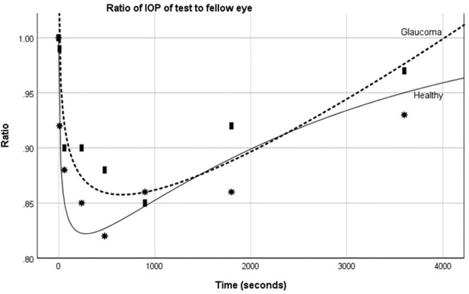
Methods: Intraocular pressure was measured using the Icare rebound tonometer (ICRT; Icare Finland Oy) and the biomechanically corrected IOP (bIOP) using the CST. Intraocular pressure was measured at baseline with ICRT, followed by a CST measurement in one eye with the fellow eye acting as a control. Icare measurements were repeated at 10 seconds and 1, 2, 4, 8, 15, 30, and 60 minutes in both eyes. The ratio of test eye IOP to fellow eye IOP was used to control for intrasubject variation.
Main outcome measures: Intraocular pressure change following Corneal Visualization Scheimflug Technology tonometry.
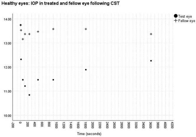
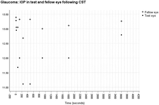
Results: Forty participants (mean age, 54.09 ± 20.08 years) were included comprising 20 patients with POAG and 20 patients with no ocular abnormalities other than cataract. Mean central corneal thickness was similar in those without POAG (547.4 ± 55.05 μm) and with POAG (520.22 ± 37.59 μm; P = 0.14). No significant change was found in IOP measured with the ICRT in the fellow eye versus the 1-hour period in either the healthy (P = 0.87) or POAG (P = 0.92) group. Significant changes were found in IOP after CST measurement for both healthy (P < 0.01) and glaucomatous (P < 0.01) eyes. After the CST measurement, the IOP reduced continuously from a mean of 13.75 mmHg to 10.84 mmHg at 4 minutes for healthy eyes and from 13.28 mmHg to 11.11 mmHg at 8 minutes for glaucomatous eyes before approaching (83% for healthy eyes and 92% POAG eyes) the pre-CST measurement at 1 hour.
Conclusions: Corneal Visualization Scheimpflug Technology tonometry causes a significant reduction in IOP in both glaucomatous and healthy eyes that lasts for at least 1 hour afterward.
Keywords: Biomechanics; CCT, central corneal thickness; CST; CST, Corneal Visualization Scheimpflug Technology tonometry; GAT, Goldmann applanation tonometry; Glaucoma; ICRT, Icare rebound tonometer; IOP, intraocular pressure; Intraocular pressure; POAG, primary open-angle glaucoma; bIOP, biomechanically corrected intraocular pressure.
© 2021 by the American Academy of Ophthalmology.
Figures
Similar articles
-
Intraocular pressure measurement and association with corneal biomechanics in patients underwent Descemet's stripping with endothelial keratoplasty: a comparative study.Front Med (Lausanne). 2024 Jul 12;11:1384694. doi: 10.3389/fmed.2024.1384694. eCollection 2024. Front Med (Lausanne). 2024. PMID: 39071083 Free PMC article.
-
[Pachymetry and intraocular pressure measurement by corneal visualization Scheimpflug technology (Corvis ST): A clinical comparison to the gold standard].Ophthalmologe. 2015 Sep;112(9):770-7. doi: 10.1007/s00347-014-3188-8. Ophthalmologe. 2015. PMID: 25501931 German.
-
Analysis of the influence of corneal properties and densitometry on applanation and rebound tonometry in primary open angle glaucoma.J Fr Ophtalmol. 2023 Mar;46(3):249-257. doi: 10.1016/j.jfo.2022.08.013. Epub 2023 Feb 2. J Fr Ophtalmol. 2023. PMID: 36739259
-
24-hour Intraocular pressure monitoring: the way ahead.Rom J Ophthalmol. 2019 Oct-Dec;63(4):315-320. Rom J Ophthalmol. 2019. PMID: 31915728 Free PMC article. Review.
-
Corneal Biomechanical Assessment with Ultra-High-Speed Scheimpflug Imaging During Non-Contact Tonometry: A Prospective Review.Clin Ophthalmol. 2021 Apr 6;15:1409-1423. doi: 10.2147/OPTH.S301179. eCollection 2021. Clin Ophthalmol. 2021. PMID: 33854295 Free PMC article. Review.
Cited by
-
Comparison of efficacy and safety between gonioscopy-assisted transluminal trabeculotomy and trabeculectomy for primary open-angle glaucoma treatment: a retrospective cohort study.BMC Ophthalmol. 2024 Dec 20;24(1):533. doi: 10.1186/s12886-024-03798-8. BMC Ophthalmol. 2024. PMID: 39702075 Free PMC article.
-
Short-term digital ocular massage may weaken corneal biomechanics.Front Bioeng Biotechnol. 2025 Jun 18;13:1582973. doi: 10.3389/fbioe.2025.1582973. eCollection 2025. Front Bioeng Biotechnol. 2025. PMID: 40606911 Free PMC article.
-
Diagnosis of Subclinical Keratoconus with a Combined Model of Biomechanical and Topographic Parameters.J Clin Med. 2021 Jun 22;10(13):2746. doi: 10.3390/jcm10132746. J Clin Med. 2021. PMID: 34206580 Free PMC article.
-
Longitudinal Analysis of Corneal Biomechanics of Suspect Keratoconus: A Prospective Case-Control Study.Bioengineering (Basel). 2024 Apr 25;11(5):420. doi: 10.3390/bioengineering11050420. Bioengineering (Basel). 2024. PMID: 38790289 Free PMC article.
-
Aerosolization ocular surface microorganisms accumulation effect during non-contact tonometer measurements.BMC Ophthalmol. 2024 Sep 3;24(1):392. doi: 10.1186/s12886-024-03664-7. BMC Ophthalmol. 2024. PMID: 39227827 Free PMC article.
References
-
- Tham Y.-C., Li X., Wong T.Y., et al. Global prevalence of glaucoma and projections of glaucoma burden through 2040: a systematic review and meta-analysis. Ophthalmology. 2014;121:2081–2090. - PubMed
LinkOut - more resources
Full Text Sources
