

Volumetric Measurement of Peripapillary Hyperreflective Ovoid Masslike Structures in Patients with Optic Disc Drusen
- PMID: 36246173
- PMCID: PMC9562331
- DOI: 10.1016/j.xops.2021.100096
Volumetric Measurement of Peripapillary Hyperreflective Ovoid Masslike Structures in Patients with Optic Disc Drusen
Abstract
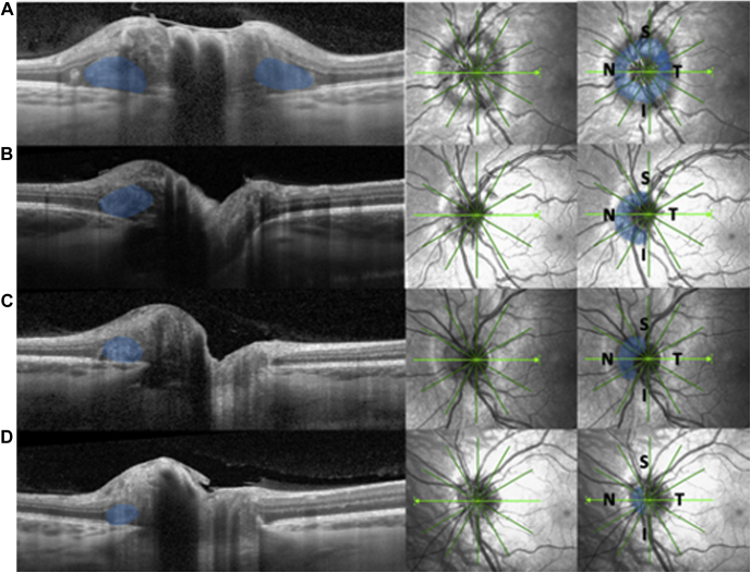
Purpose: To develop a method to determine the volume of peripapillary hyperreflective ovoid masslike structures (PHOMS) and to examine the correlation between PHOMS and anatomic optic nerve head characteristics in a large cohort of patients with optic disc drusen (ODD).
Design: Retrospective, observational study of patients with ODD.
Participants: Patients with ODD seen in a 3-year period.
Methods: We determined the prevalence of PHOMS. We then developed a method to calculate the volume of PHOMS and measured this in all patients where radial scans on OCT were available. We analyzed the correlation between PHOMS volume and patient age, size of Bruch's membrane opening (BMO), ODD visibility, and anatomic location of ODD in the optic nerve.
Main outcome measures: Prevalence and characteristics of PHOMS in patients with ODD.
Results: In 247 (77%) eyes with ODD, PHOMS were found. Among these, 80% were in the first decade of life, 87% were in the second decade, 89% were in the third decade, 85% were in the fourth decade, 74% were in the fifth decade, 73% were in the sixth decade, 58% were in the seventh decade, 40% were in the eighth decade, and 0% were in the ninth decade. The ophthalmoscopic visibility of ODD increased with age. The volume of PHOMS decreased with age, but with no correlation to the size of BMO. The median volume of PHOMS was 0.27 mm3 (interquartile range [IQR], 0.13-0.49 mm3). Predominantly, PHOMS were observed in the nasal peripapillary area (87.5% nasal, 78.5% superior, 67% inferior, and 63.5% temporal).
Conclusions: In patients with ODD, PHOMS are seen frequently, with the highest prevalence in younger individuals. The volume of PHOMS decreases with age, and PHOMS are seen more frequently in patients with superficial ODD.
Keywords: BMO, Bruch’s membrane opening; EDI, enhanced depth imaging; IQR, interquartile range; NAAION, nonarteritic anterior ischemic optic neuropathy; OCT; ODD, optic disc drusen; Optic disc drusen; Optic nerve anatomy; Optic nerve head drusen; PHOMS; PHOMS, peripapillary hyperreflective ovoid masslike structure(s); Peripapillary hyperreflective ovoid masslike structure; Volumetric measurement.
© 2021 by the American Academy of Ophthalmology.
Figures


References
-
- Spaide R.F., Koizumi H., Pozzoni M.C. Enhanced depth imaging spectral-domain optical coherence tomography. Am J Ophthalmol. 2008;146:496–500. - PubMed
-
- Fraser J.A., Hamann S.A. 360-degree peripapillary hyper-reflective ovoid mass-like structure (PHOMS) Can J Ophthalmol. 2021;56:146. - PubMed
-
- Malmqvist L., Bursztyn L., Costello F., et al. Peripapillary hyperreflective ovoid mass-like structures: is it optic disc drusen or not? Response. J Neuroophthalmol. 2018;38:568–570. - PubMed
-
- Malmqvist L., Bursztyn L., Costello F., et al. The Optic Disc Drusen Studies Consortium recommendations for diagnosis of optic disc drusen using optical coherence tomography. J Neuroophthalmol. 2018;38:299–307. - PubMed
-
- Traber G.L., Weber K.P., Sabah M., et al. Enhanced depth imaging optical coherence tomography of optic nerve head drusen: a comparison of cases with and without visual field loss. Ophthalmology. 2017;124:66–73. - PubMed
LinkOut - more resources
Full Text Sources
