

Evaluation of a droplet digital PCR assay for quantification of Mycobacterium avium subsp. paratuberculosis DNA in whole-blood and fecal samples from MAP-infected Holstein cattle
- PMID: 36246323
- PMCID: PMC9563315
- DOI: 10.3389/fvets.2022.944189
Evaluation of a droplet digital PCR assay for quantification of Mycobacterium avium subsp. paratuberculosis DNA in whole-blood and fecal samples from MAP-infected Holstein cattle
Abstract
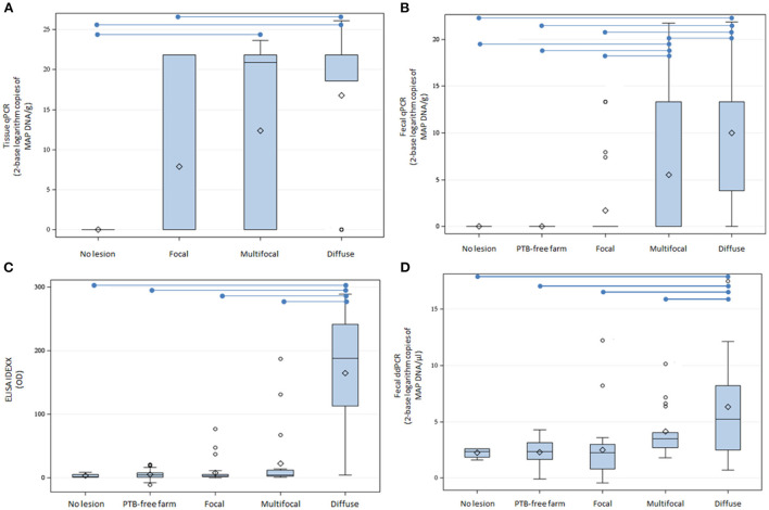
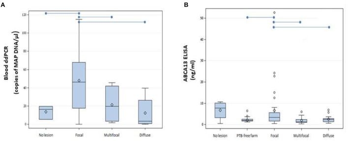
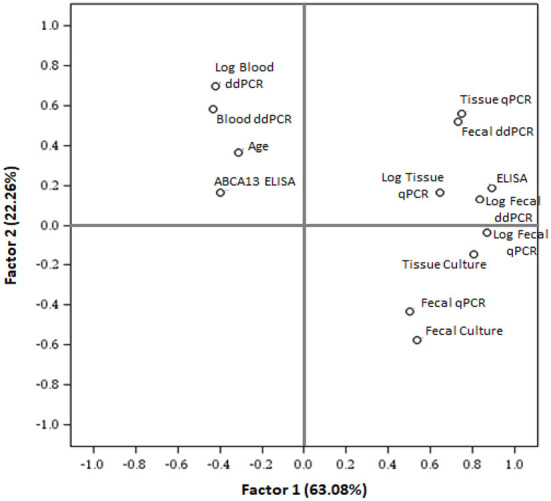
Bovine paratuberculosis (PTB) is an infectious disease that affects ruminants worldwide and is a burden on the dairy industry. PTB control measures include culling of Mycobacterium avium subsp. paratuberculosis (MAP)-infected animals from the herd and the enhancement of farm-biosecurity measures. Diagnostics tools for the direct detection of MAP are fecal real-time qPCR and bacteriological culture, the last one being considered the gold standard. However, both show limitations for detecting subclinical MAP-infected cattle with low bacterial load in feces and gut tissues. Droplet digital polymerase chain reaction (ddPCR) is a third-generation PCR method that shows high reproducibility for the quantification of low DNA copy numbers. The objective of this study was to design a ddPCR assay to detect and quantify a fragment of the F57 MAP-specific sequence in samples of naturally MAP-infected Holstein cattle. DNA was isolated from whole-blood and fecal samples from control cows with a negative ELISA and qPCR result (N = 75) and from cows with PTB-associated focal (N = 32), multifocal (N = 21), and diffuse lesions (N = 17) in gut tissues. After ddPCR, the DNA extracted from fecal samples of cows with diffuse lesions showed higher mean copies per microliter (13,791.2 copies/μl) than samples from cows with multifocal lesions (78.8 copies/μl), focal lesions (177.1 copies/μl) or control cows (4.8 copies/μl) (P ≤ 0.05). Significant differences in mean DNA copies/μl were also observed in the blood samples from cows with focal lesions (47.7 copies/μl) when compared with cows with multifocal and diffuse lesions; 18.1 and 12.4 copies/μl, respectively. Using a principal component analysis, the results of the fecal ddPCR clustered together with the results of a commercial ELISA for the specific detection of MAP antibodies, fecal and tissue qPCR, and bacteriological culture results. In contrast, blood ddPCR results clustered together with the results of an ELISA for the detection of a biomarker of subclinical PTB, the ABCA13 transporter. Blood ddPCR was the most sensitive tool (sensitivity 71%, specificity 100%) of all the quantitative methods used in the study for the detection of subclinical cows with focal lesions.
Keywords: blood; droplet digital PCR; feces; molecular diagnosis; paratuberculosis.
Copyright © 2022 Badia-Bringué, Canive, Casais, Blanco-Vázquez, Amado, Iglesias, González, Bascones, Juste and Alonso-Hearn.
Conflict of interest statement
The authors declare that the research was conducted in the absence of any commercial or financial relationships that could be construed as a potential conflict of interest.
Figures
References
-
- Bermudez LE, Petrofsky M, Sommer S, Barletta RG. Peyer's patch-deficient mice demonstrate that Mycobacterium avium subsp. paratuberculosis translocates across the mucosal barrier via both M cells and enterocytes but has inefficient dissemination. Infect Immun. (2010) 78:3570–7. 10.1128/IAI.01411-09 - DOI - PMC - PubMed
LinkOut - more resources
Full Text Sources