. 2021 Dec 6;1(4):100087.
doi: 10.1016/j.xops.2021.100087. eCollection 2021 Dec.

Genetic Risk in Families with Age-Related Macular Degeneration

Affiliations

Genetic Risk in Families with Age-Related Macular Degeneration

Anita de Breuk et al. Ophthalmol Sci. .

Abstract

Purpose: To determine the contribution of common and rare genetic risk variants in families with age-related macular degeneration (AMD).

Design: Case-control study.

Participants: A family cohort (355 affected and 342 unaffected family members from 144 families with AMD) and an unrelated case-control cohort (1078 patients, 952 controls), recruited from the European Genetic Database.

Methods: Genetic data of both cohorts were filtered for carriership of rare genetic variants in the coding and splice-site regions of the complement factor H (CFH) and complement factor I (CFI) genes, and 52 AMD-associated variants were extracted for calculation of genetic risk scores (GRS). To compare GRSs between familial and nonfamilial rare CFH and CFI variant carriers and noncarriers and between AMD disease stages, we performed a 2-way analysis of variance, with Bonferroni correction for multiple testing. Within families with AMD carrying rare CFH and CFI variants, we analyzed segregation patterns by calculating the proportion of affected among carriers.

Main outcome measures: GRSs and segregation of rare CFH and CFI variants.

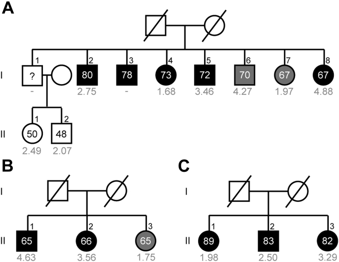
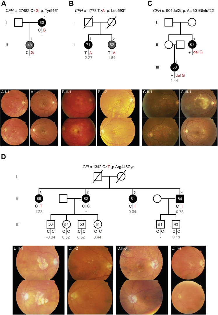
Results: We observed higher GRSs in familial versus nonfamilial individuals without rare CFH and CFI variants: mean GRS, 1.76 (standard error [SE], 0.08) versus 0.83 (SE, 0.03; P < 0.001). In 51 of 144 families (35.4%), rare CFH and CFI variants were identified. Within the AMD family cohort, carriers of rare CFH and CFI variants showed lower GRSs compared with noncarriers (mean GRS, 1.05 [SE, 0.23] vs. 1.76 [SE, 0.08]; P = 0.02). The proportion of affected family members with a high GRS was 57.3% (176/307). Of the affected family members with a low or intermediate GRS, 40.0% carried rare CFH or CFI variants. Among carriers of 11 rare CFH or CFI variants, the proportion affected by AMD was more than 75%.

Conclusions: Genetic risk in families with AMD often is attributed to high GRSs based on common variants. However, in part of the families with a low or intermediate GRS, rare CFH and CFI variants contributed to disease development. We recommend computing GRSs and sequencing the CFH and CFI genes in families with AMD, in particular in the light of ongoing gene-specific clinical trials.

Keywords: AMD, age-related macular degeneration; Age-related macular degeneration; CCP, complement control protein; CFH, complement factor H; CFI, complement factor I; CI, confidence interval; CIRCL, Cologne Image Reading Center and Laboratory; CNV, choroidal neovascularization; Complement factor H; Complement factor I; Complement system; GA, geographic atrophy; GRS, genetic risk score; GWAS, genome-wide association study; Genetic risk score; IQR, interquartile range; RC, Rotterdam Classification; SE, standard error.


Figures

Figure 1
Figure 1
Graphs showing the genetic risk score in familial age-related macular degeneration (AMD) and unrelated individuals. The y-axis represents the estimated marginal mean genetic risk score. The x-axis represents the AMD disease stages. The family cohort is indicated in red, and the unrelated case-control cohort is indicated in blue. Dotted lines represent rare CFH and CFI variant carriers, and continuous lines represent CFH and CFI noncarriers. Error bars are ±1 standard error. Numbers within the figures indicate the estimated marginal mean genetic risk scores with corresponding standard errors. A 2-way analysis of variance was performed to compare genetic risk scores among the 4 groups and among the AMD disease stages. A Bonferroni correction for multiple testing was applied for all pairwise comparisons (BD). A, Genetic risk score for all categories and AMD disease stages. B, Pairwise comparisons of AMD disease stages and group categories. C, Pairwise comparisons within the family cohort. D, Pairwise comparisons within the case-control cohort. ∗P < 0.05; ∗∗P < 0.01; ∗∗∗P < 0.001. int = intermediate; ns = not significant; RV = rare variant.
Figure 2
Figure 2
Pedigrees of 3 families with age-related macular degeneration (AMD) without rare CFH or CFI variants and a high median genetic risk score (A-C). Individuals affected by early or intermediate AMD are indicated in grey, and individuals affected by advanced AMD are indicated in black. Age at examination and the genetic risk score (in grey) are given for each individual.
Figure 3
Figure 3
Pedigrees of 2 families with age-related macular degeneration (AMD) with rare CFH variants and a low median genetic risk score (A-B). Individuals affected by early or intermediate AMD are indicated in grey, and individuals affected by advanced AMD are indicated in black. Individual III.2 and III.3 from family B are affected by peripheral cuticular drusen (B, indicated in light grey). Age at examination and the genetic risk score (in grey) are given for each individual. A rare variant in the CFH gene was identified in family A, c.2572T→A, p.Trp858Arg (A), and in family B, c.1222C→T, p.Gln408∗ (B). Carriers of the risk allele are indicated in red. aGenetic risk score of individual III.3 from family A (A) and individual II.2 from family B (B) is incomplete because they have a missing genotype in 2 of the 5 major risk alleles.
Figure 4
Figure 4
Pedigrees with accompanying color fundus photographs of 4 families with age-related macular degeneration (AMD) carrying rare CFH and CFI variants that segregate with AMD phenotype. Individuals affected by early or intermediate AMD are indicated in grey, and individuals affected by advanced AMD are indicated in black. Age at examination and the genetic risk score (in grey) are given for each individual. Carriers of the risk allele are indicated in red. A, Family carrying the rare CFH p.Tyr916∗ variant. B, Family carrying the rare CFH p.Leu593∗ variant. C, Family carrying the rare CFH p.Ala301Glnfs∗22 variant. D, Family carrying the rare CFI p.Arg448Cys variant.
Figure 5
Figure 5
Graph showing the segregation patterns and genetic risk scores (GRSs) of carriers of rare CFH and CFI variants that manifest age-related macular degeneration (AMD). The y-axis represents the median genetic risk score per variant, and the x-axis represents the number of affected carriers divided by the total number of carriers (percentage). The variants are color coded according to the GRS category: grey dots correspond to the low GRS category (≤0.220), orange dots correspond to the intermediate GRS category (0.221–1.407), and red dots correspond to the high GRS category (≥1.408).

References

    1. Colijn J.M., Buitendijk G.H.S., Prokofyeva E., et al. Prevalence of age-related macular degeneration in Europe: the past and the future. Ophthalmology. 2017;124(12):1753–1763. - PMC - PubMed
    1. Flaxman S.R., Bourne R.R.A., Resnikoff S., et al. Global causes of blindness and distance vision impairment 1990–2020: a systematic review and meta-analysis. Lancet Glob Health. 2017;5(12):e1221–e1234. - PubMed
    1. Meyers S.M., Greene T., Gutman F.A. A twin study of age-related macular degeneration. Am J Ophthalmol. 1995;120(6):757–766. - PubMed
    1. Seddon J.M., Cote J., Page W.F., et al. The US twin study of age-related macular degeneration: relative roles of genetic and environmental influences. Arch Ophthalmol. 2005;123(3):321–327. - PubMed
    1. Klaver C.C., Wolfs R.C., Assink J.J., et al. Genetic risk of age-related maculopathy. Population-based familial aggregation study. Arch Ophthalmol. 1998;116(12):1646–1651. - PubMed

LinkOut - more resources