. 2022 Sep 15;14(9):6067-6081.
eCollection 2022.

Discoidin domain receptor 2 expression increases phagocytotic capacity in sertoli cells of sertoli cell-only syndrome testes

Affiliations

Discoidin domain receptor 2 expression increases phagocytotic capacity in sertoli cells of sertoli cell-only syndrome testes

Ying Chen et al. Am J Transl Res. .

Abstract

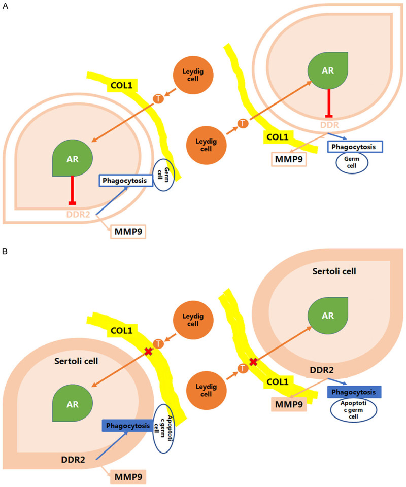
Discoidin domain receptor 2 (DDR2) belongs to the receptor tyrosine kinase (RTK) family, other RTKs have been reported to regulate phagocytic function of Sertoli cells (SCs), yet little is known about the function of DDR2 in Sertoli cells. In the present study, we aim to explore the function and mechanism of ectopic discoidin domain receptor 2 (DDR2) expression in Sertoli cells of Sertoli cell-only syndrome (SCOS) testes. We found that discoidin domain receptor 2 (DDR2) was absent in Sertoli cells of normal testis but was expressed in Sertoli cells of SCOS testes. This Sertoli cell DDR2 expression was induced by impaired androgen receptor (AR) signaling, but was inhibited by increased AR signaling from testosterone administration. The Sertoli cell DDR2 expression led to an increase in phagocytosis through up-regulation of Scavenger receptor class B member 1 (SR-BI) levels. However, loss of DDR2 by knock-out or knock-down weakened the phagocytotic capacity of Sertoli cells. Furthermore, the expression of DDR2 in Sertoli cells activated matrix metallopeptidase 9 (MMP-9) to consume abnormal collagen increase in seminiferous tubules which was responsible for the block of testosterone transportation and AR loss and to compensate for the impaired blood-testis-barrier (BTB). Our data suggest that the AR/DDR2 cascade may serve as a negative feedback mechanism to help compensate for the homeostasis of seminiferous epithelium in SCOS testis.

Keywords: Sertoli cell-only syndrome (SCOS); androgen receptor (AR); discoidin domain receptor 2 (DDR2); phagocytosis; sertoli cell.


Conflict of interest statement

None.

Figures

Figure 1
Figure 1
Histology and immunohistochemistry of Sertoli cell only syndrome testis (40X). H&E staining of control testis (A) and Sertoli cell only syndrome (SCOS) testis (B). COL1 expression in control testis (C) and SCOS testis (D). COL1 immunoactivity was found in basal membrane of control testis and Sertoli cell only syndrome testis, the COL1 immunoactivity in SCOS testis was stronger than control testis. Arrow head indicates basal membrane. (E, F) DDR2 immunohistochemical staining in control human testis and SCOS patient testis. DDR2 was exclusively expressed in Leydig cells in control testis but was both expressed in Leydig cells and Sertoli cells in SCOS testis. Arrow indicates Leydig cell and arrow head represent Sertoli cell. Bar =50 μm.
Figure 2
Figure 2
AR and DDR2 expression in human control testis and SCOS testis (40X). AR and DDR2 co-localization in human control testis (A, C, E, G). No DDR2 (C) immunoreactivity was found in seminiferous tubules of control testis while AR (E) was found located in Sertoli cells. AR and DDR2 co-localization in SCOS testis (B, D, F, H). Strong DDR2 (D) expression was found in Sertoli cell of SCOS tubules while AR (F) expression was lost in SCOS seminiferous tubules. AR was stained with green, while DDR2 was stained with red. DAPI was stained with blue indicating nuclear. Arrow heads indicate Sertoli cells. Bar =50 μm.
Figure 3
Figure 3
DDR2 expression was induced in Sertoli cell by decrease of AR signaling (A-F, 40X). DDR2 expression in Sertoli cell of seminiferous tubules after EDS 0 h (A), 6 h (B), 12 h (C), 24 h (D), 48 h (E) and 72 h (F). DDR2 expression was negative in EDS 0h testis (A) but became positive in EDS treated 12 h (C) and 24 h (D) testis. Arrow head indicates DDR2 positive Sertoli cell. Bar =50 μm. (G) Typical western blot results of DDR2 and AR expression in TM4 Sertoli cells with different concentration T treatment. AR levels in TM4 cells increased with elevated testosterone levels. DDR2 expression was found in TM4 cells but was inhibited by 100 nmol/L testosterone treatment. (H) Flutamide pre-treatment reverses the testosterone inhibition to DDR2.
Figure 4
Figure 4
Phagocytotic capacity increased in DDR2 over-expressed Sertoli cell but decreased in DDR2 knock-down Sertoli cell. (A) Staining of rodamin labeled RBs phagocytosis in control and DDR2 over-expressed Sertoli cells. Bar =50 μm. The magnification of (A) was 40X. (B) DDR2 mRNA level in control and DDR2 over-expressed Sertoli cells. The mRNA level of DDR2 in over-expressed group was 2.6 folds higher than control group. (C) Number of phagocytotic RBs in control and DDR2 over-expressed Sertoli cells. The number of phagocytotic RBs in DDR2 over-expressed Sertoli cells was 3 folds higher than control group. (D) SR-BI mRNA level in control and DDR2 over-expressed Sertoli cells. The SR-BI mRNA level in DDR2 over-expressed Sertoli cells was 2 folds higher than control group. (E) Staining of rodamin labeled RBs phagocytosis in negative control Sertoli cells and DDR2 knock-down Sertoli cells. Bar =50 μm. The magnification of (E) was 40X. (F) DDR2 mRNA level in negative control and DDR2 knock-down Sertoli cells. The DDR2 mRNA level in knock-down Sertoli cells was 40% of control group. (G) Number of phagocytotic RBs in negative control and DDR2 knock-down Sertoli cells. The number of phagocytotic RBs in DDR2 knock-down Sertoli cells was less than 50% of control group. (H) SR-BI mRNA level in negative control and DDR2 knock-down Sertoli cells. The SR-BI mRNA level in DDR2 knock-down Sertoli cells was less than 50% of control group.
Figure 5
Figure 5
Phagocytosis capacity decreased in Sertoli cell of DDR2 deficiency mouse testis. (A, B) DDR2 immunostaining in wild type (A) and DDR2slie/slie (B) mouse testis. Bar =50 μm. (C, D) PAS staining of wild type (C) and DDR2slie/slie (D) testicular section. Arrow head indicates the residual body. Bar =50 μm. (E, F) Oil red O staining of wild type (E) and DDR2slie/slie (F) testicular section. Arrow heads represents the lipid drop in Sertoli cell. Bar =50 μm. (G) RBs number in wild type and DDR2slie/slie mouse testis. The average RBs number in tubule at stage VIII of DDR2slie/slie mouse testis was 2 folds higher than control group. The magnification was 40X. (H) Number of lipid drop in per Sertoli cell of wild type and DDR2slie/slie mouse testis. The number of lipid drop in per Sertoli cell of DDR2slie/slie mouse testis was 2.6 folds higher than control testis. (I) SR-BI mRNA level in wild type and DDR2slie/slie testis. The SR-BI mRNA levels in DDR2slie/slie testis were less than 50% of control testis.
Figure 6
Figure 6
Up-regulated DDR2 expression in SCs of SCOS testis activated MMP-9 to consume the abnormal increased collagen and to compensate the impaired blood-testis-barrier. (A-D) Laser captured dissection of Sertoli cell from control testis (A and B) and SCOS testis (C and D). Bar =50 μm. The magnification of was 40X. (E) SR-BI mRNA level in control and SCOS testicular Sertoli cell. The SR-BI mRNA level in SCOS testicular Sertoli cell was 1.8 folds higher than control group. (F) N-cadherin mRNA level in control and SCOS testicular Sertoli cell. The N-cadherin mRNA level in SCOS testicular Sertoli cell was 3.1 folds higher than control group. (G) Occludin mRNA level in control and SCOS testicular Sertoli cell. The occludin mRNA level in SCOS testicular Sertoli cell was 3.3 folds higher than control group. (H) MMP-9 mRNA level in control and SCOS testicular Sertoli cell. The MMP-9 mRNA level in SCOS testicular Sertoli cell was 3 folds higher than control group. (I) Immunoblot of DDR2 and MMP-9 expression in TM4 Sertoli cells with different dose T administration plus collagen I pre-treatment. (J) Immunoblot of DDR2 and MMP-9 expression in DDR2 over-expressed TM4 Sertoli cells with 100 nM T administration plus collagen I pre-treatment.
Figure 7
Figure 7
Scheme about the signaling and the role of up-regulated DDR2 in control human and SCOS testis. A. In control testis, the Sertoli cell DDR2 expression was inhibited by androgen receptor (AR) activated by Leydig cell produced testosterone. B. In SCOS testis, the increased collagen in basal membrane impaired the testosterone transportation and led to AR inactivation. The AR inactivation induced DDR2 expression, which promoting phagocytosis and activating MMP-9.

References

    1. Kierszenbaum AL. Mammalian spermatogenesis in vivo and in vitro: a partnership of spermatogenic and somatic cell lineages. Endocr Rev. 1994;15:116–134. - PubMed
    1. Rodriguez I, Ody C, Araki K, Garcia I, Vassalli P. An early and massive wave of germinal cell apoptosis is required for the development of functional spermatogenesis. EMBO J. 1997;16:2262–2270. - PMC - PubMed
    1. de Rooij DG, Grootegoed JA. Spermatogonial stem cells. Curr Opin Cell Biol. 1998;10:694–701. - PubMed
    1. Billig H, Furuta I, Rivier C, Tapanainen J, Parvinen M, Hsueh AJ. Apoptosis in testis germ cells: developmental changes in gonadotropin dependence and localization to selective tubule stages. Endocrinology. 1995;136:5–12. - PubMed
    1. Bartke A. Apoptosis of male germ cells, a generalized or a cell type-specific phenomenon? Endocrinology. 1995;136:3–4. - PubMed

LinkOut - more resources