

Clinical value and feasibility of CT pulmonary angiography with personalized injection of contrast agent in pulmonary embolism
- PMID: 36247283
- PMCID: PMC9556491
Clinical value and feasibility of CT pulmonary angiography with personalized injection of contrast agent in pulmonary embolism
Abstract
Objective: To determine the clinical value and feasibility of CT pulmonary angiography (CTPA) with personalized injection of contrast agent in pulmonary embolism (PE).
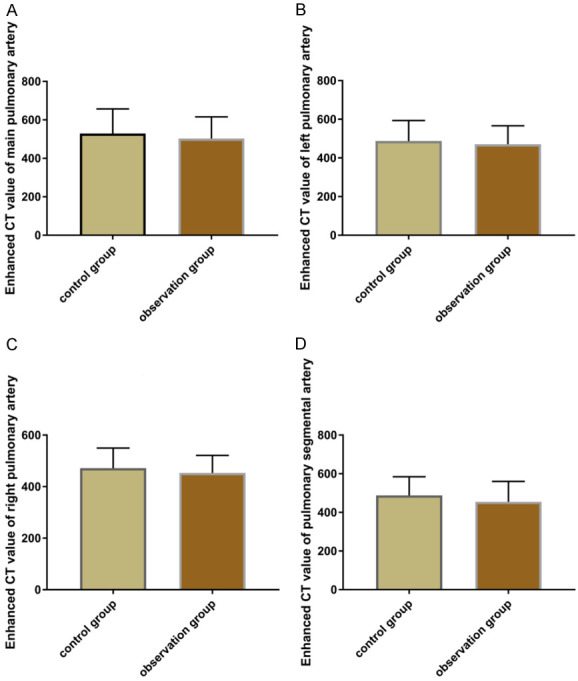
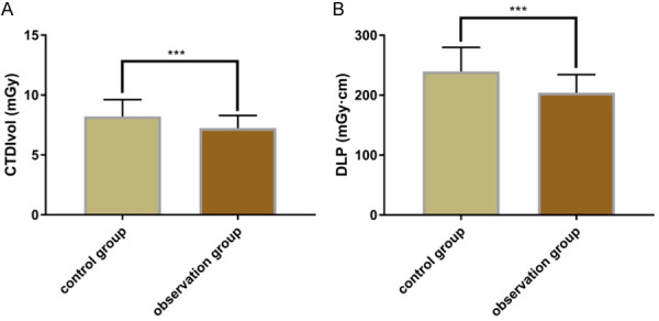
Methods: In the present retrospective study, 130 patients who underwent CTPA examination in our hospital from June 2019 to May 2020 were evaluated. Among them, 67 cases were detected by CTPA with personalized injection of contrast agent as the observation group (Obs group), and 63 cases were detected by CTPA with bolus-tracking (BT) as the control group (Con group). The specificity, sensitivity and accuracy of the detection in the two groups were compared. The image quality score and superior vena cava artifact score of the two diagnostic methods were compared. Additionally, the volumetric CT dose index (CTDIvol) and dose length product (DLP) of the two groups were compared.
Results: The Obs group yielded a significantly higher specificity in diagnosing PE than the Con group (P<0.05), but there were no significant differences between the two groups in the sensitivity and accuracy (P>0.05). The image quality score and superior vena cava artifact score of the two groups were not significantly different (P>0.05), and the Obs group showed significantly lower CTDIvol and DLP than the Con group (P<0.05).
Conclusion: CTPA with personalized injection of contrast agent has good diagnostic value for PE, with good imaging effect and safe profile, and has a lower radiation dose requirement.
Keywords: CTPA; Personalized injection of contrast agent; diagnose; pulmonary artery embolism.
AJTR Copyright © 2022.
Conflict of interest statement
None.
Figures
Similar articles
-
Feasibility of a Single Contrast Bolus High-Pitch Pulmonary CT Angiography Protocol Followed by Low-Dose Retrospectively ECG-Gated Cardiac CT in Patients with Suspected Pulmonary Embolism.Rofo. 2018 Jun;190(6):542-550. doi: 10.1055/s-0044-100725. Epub 2018 Feb 1. Rofo. 2018. PMID: 29390229 English.
-
[Application of Low Concentration Contrast Agent Combined Double Low Dose in CT Pulmonary Angiography for Pulmonary Embolism].Sichuan Da Xue Xue Bao Yi Xue Ban. 2018 Mar;49(2):239-242. Sichuan Da Xue Xue Bao Yi Xue Ban. 2018. PMID: 29737068 Clinical Trial. Chinese.
-
An optimized test bolus for computed tomography pulmonary angiography and its application at 80 kV with 10 ml contrast agent.Sci Rep. 2020 Jun 23;10(1):10208. doi: 10.1038/s41598-020-67145-9. Sci Rep. 2020. PMID: 32576901 Free PMC article.
-
[The role of CT pulmonary angiography in the diagnosis and prognosis of pulmonary embolism and correlation with blood gas values].Zhonghua Jie He He Hu Xi Za Zhi. 2012 Oct;35(10):770-4. Zhonghua Jie He He Hu Xi Za Zhi. 2012. PMID: 23289996 Chinese.
-
A Systematic Review of Double Low-dose CT Pulmonary Angiography in Pulmonary Embolism.Curr Med Imaging Rev. 2019;15(5):453-460. doi: 10.2174/1573405614666180813120619. Curr Med Imaging Rev. 2019. PMID: 32008552
References
-
- Ibrahim WH, Al-Shokri SD, Hussein MS, Kamel A, Abu Afifeh LM, Karuppasamy G, Parambil JV, Elasad FM, Abdelghani MS, Abdellah A, Faris ME. Saddle versus non-saddle pulmonary embolism: differences in the clinical, echocardiographic, and outcome characteristics. Libyan J Med. 2022;17:2044597. - PMC - PubMed
-
- Nagraj S, Li WJ, Zamora C, Barakakis PA, Kokkinidis DG. Pharmacological and interventional management of pulmonary embolism: where do we stand? Future Cardiol. 2022;18:191–206. - PubMed
-
- Ekici M, Ekici A, Ileri S. Chronic CT features in PE patients with co-existing DVT. Am J Emerg Med. 2021;46:126–131. - PubMed
-
- Loftus JR, Hu ZX, Morin BR, Hobbs SK, Francis CW, Khorana AA, Rubens DJ, Kaproth-Joslin KA. Vascular imaging in the asymptomatic high-risk cancer population: a role for thrombosis screening and therapy management. J Ultrasound Med. 2022;41:225–236. - PubMed
LinkOut - more resources
Full Text Sources