. 2022 Sep 14;8(7):FSO811.
doi: 10.2144/fsoa-2022-0005. eCollection 2022 Aug.

The promising role of hypoxia-resistant insulin-producing cells in ameliorating diabetes mellitus in vivo

Affiliations

The promising role of hypoxia-resistant insulin-producing cells in ameliorating diabetes mellitus in vivo

Hanaa H Ahmed et al. Future Sci OA. .

Abstract

Aim: This study aimed to evaluate the efficacy of hypoxia-persistent insulin-producing cells (IPCs) against diabetes in vivo.

Materials & methods: Mesenchymal stem cells (MSCs) differentiation into IPCs in the presence of Se/Ti (III) or CeO2 nanomaterials. IPCs were subjected to hypoxia and hypoxia genes were analyzed. PKH-26-labeled IPCs were infused in diabetic rats to evaluate their anti-diabetic potential.

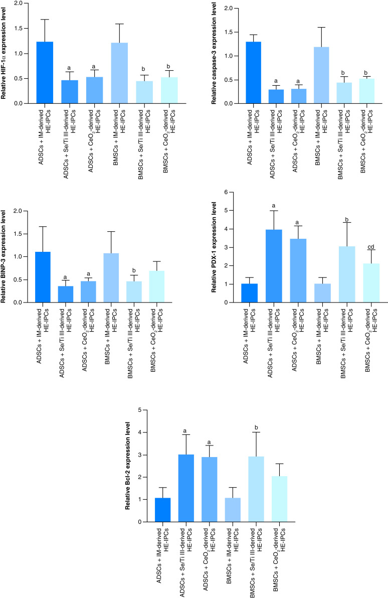
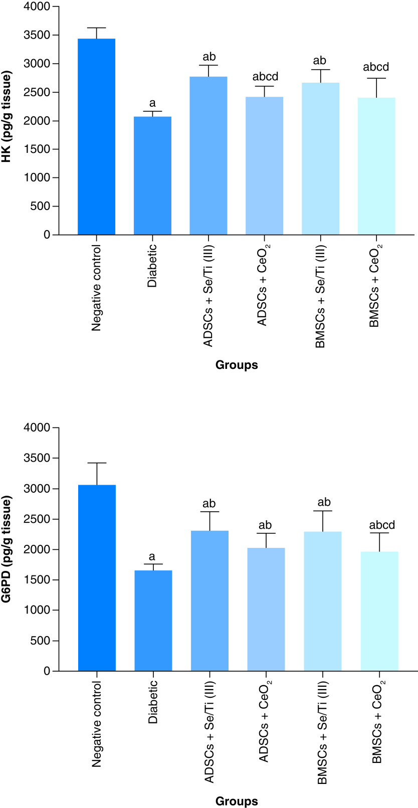
Results: MSCs were differentiated into functional IPCs. IPCs exhibited overexpression of anti-apoptotic genes and down-expression of hypoxia and apoptotic genes. IPCs implantation elicited glucose depletion and elevated insulin, HK and G6PD levels. They provoked VEGF and PDX-1 upregulation and HIF-1α and Caspase-3 down-regulation. IPCs transplantation ameliorated the destabilization of pancreatic tissue architecture.

Conclusion: The chosen nanomaterials were impressive in generating hypoxia-resistant IPCs that could be an inspirational strategy for curing diabetes.

Keywords: diabetes mellitus; hypoxia; insulin-producing cells; mesenchymal stem cells; nanomaterials.


Figures

Figure 1.
Figure 1.. Relative mRNA expression analysis of insulin-producing cells-related genes (Nkx2.2, Ngn-3 and PDX-1) in adipose-derived stem cells and bone marrow mesenchymal stem cells-derived insulin-producing cells.
Data are displayed as (mean ± SD) procured from four independent experiments (n = 4). (A) Significant variation at p < 0.05 relative to undifferentiated ADSCs. (B) Significant variation at p < 0.05 relative to (ADSCs + IM). (C) Significant variation at p < 0.05 relative to undifferentiated BMSCs. (D) Significant variation at p < 0.05 versus (BMSCs +IM). (E) Significant change at p < 0.05 relative to (ADSCs + Se/Ti III). (F) Significant variation at p < 0.05 relative to (BMSCs + Se/Ti III). ADSC: Adipose-derived stem cell; BMSC: Bone marrow mesenchymal stem cell; IM: Inductive media.
Figure 2.
Figure 2.. Relative gene expression analysis of hypoxia-related genes; proapoptotic genes (HIF-1α, Caspase-3, BNIP3) and anti-apoptotic genes (Bcl-2 and PDX-1) in adipose-derived stem cells and bone marrow mesenchymal stem cells-derived hypoxia exposed-insulin-producing cells.
Data are displayed as (mean ± SD) acquired from four independent experiments (n = 4). (A) Significant change at p < 0.05 relative to (ADSCs-derived HE-IPCs + IM). (B) Significant change at p < 0.05 relative to (BMSCs-derived HE-IPCs + IM). (C) Significant change at p < 0.05 relative to (ADSCs-derived HE-IPCs + Se/Ti III). (D) Significant change at p < 0.05 relative to (ADSCs-derived HE-IPCs + CeO2). ADSC: Adipose-derived stem cell; BMSC: Bone marrow mesenchymal stem cell; HE-IPC: Hypoxia exposed-insulin-producing cell; IM: Inductive media.
Figure 3.
Figure 3.. Homing of the implanted insulin-producing cells to the pancreatic tissues of different treated groups indicating the presence of red-labeled cells.
(A) Adipose-derived stem cells + Se/Ti (III). (B) Adipose-derived stem cells + CeO2. (C) Bone marrow mesenchymal stem cells + Se/Ti (III). (D) Bone marrow mesenchymal stem cells + CeO2 group.
Figure 4.
Figure 4.. Effect of hypoxia-resistant insulin-producing cells transplantation on plasma glucose and serum level.
(A) Plasma glucose level of diabetic rats. (B) Serum level of insulin of diabetic rats. Values are expressed as (means ± SD) of eight rats per group. (a) Significant difference at p < 0.05 when compared with the negative control group. (b) Significant difference at p < 0.05 when compared with the diabetic group. (c) Significant difference at p < 0.05 when compared with the ADSCs + Se/Ti (III) group. (d) Significant difference at p < 0.05 when compared with the BMSCs + Se/Ti (III) group. ADSC: Adipose-derived stem cell; BMSC: Bone marrow mesenchymal stem cell.
Figure 5.
Figure 5.. Effect of hypoxia-resistant insulin-producing cells transplantation on the hepatic carbohydrate metabolizing enzyme activities (HK and G6PD) of the diabetes-induced rats.
Values are displayed as (means ± SD) of eight rats per group. (A) Significant variation at p < 0.05 in comparison with the negative control group. (B) Significant variation at p < 0.05 in comparison with the diabetic group. (C) Significant variation at p < 0.05 in comparison with the ADSCs + Se/Ti (III) group. (D) Significant variation at p < 0.05 in comparison with the BMSCs + Se/Ti (III) group. ADSC: Adipose-derived stem cell; BMSC: Bone marrow mesenchymal stem cell.
Figure 6.
Figure 6.. Effect of implanted hypoxia-resistant insulin-producing cells on the pancreatic VEGF, PDX-1, HIF-1α and Caspase-3 gene expression patterns of the diabetic rats.
Values are represented as (means ± SD) of four rats per group. (A) Significant variation at p < 0.05 versus the negative control group. (B) Significant variation at p < 0.05 versus the diabetic group. ADSC: Adipose-derived stem cell; BMSC: Bone marrow mesenchymal stem cell.
Figure 7.
Figure 7.. Histological investigations of pancreatic tissue of different groups.
(A) Negative control rat displaying normal pancreatic architecture with active pancreatic acini. (B) Untreated diabetic rat revealing shrinkage of Langerhans islets and pancreatic acini associated with necrosis of components cells (star). (C) Diabetic rat infused with ADSCs + Se/Ti (III)-derived insulin-producing cells (IPCs) showing normal Langerhans islets and active pancreatic acini. (D) Diabetic rat implanted with adipose-derived stem cells +CeO2-derived IPCs showing shrinkage of Langerhans islets and pancreatic acini, with degeneration pyknosis and karyolysis is evident (arrow). (E) Diabetic rat infused with BMSCs + Se/Ti (III)-derived IPCs showing normal Langerhans islets (arrow) with some dilatation of blood capillaries and pancreatic ducts. (F) Diabetic rat injected with BMSCs + CeO2-derived IPCs showing shrinkage and necrosis of Langerhans islets (star) (H&E, ×400, scale bar: 3 μm).

References

    1. Yong J, Johnson JD, Arvan P, Han J, Kaufman RJ. Therapeutic opportunities for pancreatic β-cell ER stress in diabetes mellitus. Nat. Rev. Endocrinol. 17, 455–467 (2021). - PMC - PubMed
    1. Saeedi P, Petersohn I, Salpea P et al. Global and regional diabetes prevalence estimates for 2019 and projections for 2030 and 2045: Results from the International Diabetes Federation Diabetes Atlas, 9th edition. Diabetes Res. Clin. Pract. 157 (2019). . - PubMed
    1. Shelbaya S, Halawa M, Nasr M. The management of care of Egyptian patients with diabetes: a report from the International Diabetes Management Practices Study Wave 7. Med J Cairo Univ. 88(6), 1413–1421 (2020).
    1. Marcovecchio ML. Complications of acute and chronic hyperglycemia. US Endocrinol. 13(1), 17–21 (2017).
    1. Gupta R, Ghosh A, Singh AK, Misra A. Clinical considerations for patients with diabetes in times of COVID-19 epidemic. Diabetes Metab. Syndr. Clin. Res. Rev. 14, 211–212 (2020). - PMC - PubMed