

A review of the management of central retinal artery occlusion
- PMID: 36248088
- PMCID: PMC9558462
- DOI: 10.4103/2211-5056.353126
A review of the management of central retinal artery occlusion
Abstract
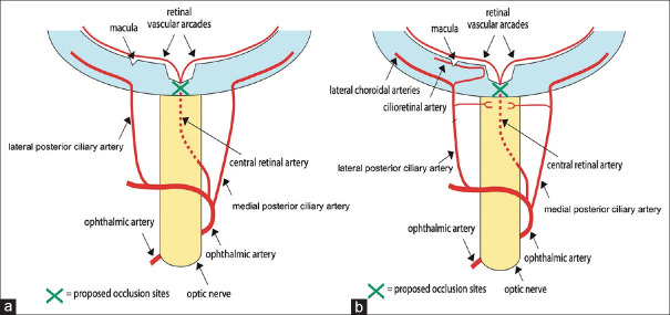
Central retinal artery occlusion (CRAO), the ocular analog of a cerebral stroke, is an ophthalmic emergency. The visual prognosis for overall spontaneous visual recovery in CRAO is low. Furthermore, the risk of future ischemic heart disease and cerebral stroke is increased due to the underlying atherosclerotic risk factors. There is currently no guideline-endorsed treatment for CRAO. This review will describe the anatomy, pathophysiology, epidemiology, and clinical features of CRAO, and investigate the current and future management strategies.
Keywords: Central retinal artery; Ischemia; occlusion; prevention; reperfusion; thrombolysis.
Copyright: © 2022 Taiwan J Ophthalmol.
Conflict of interest statement
Prof. Celia Chen, an editorial board member at Taiwan Journal of Ophthalmology, had no role in the peer review process of or decision to publish this article. The other authors decalared no conflicts of interest in writing this paper.
Figures

Similar articles
-
A Review of Current Literature on Central Retinal Artery Occlusion: Its Pathogenesis, Clinical Management, and Treatment.Cureus. 2024 Mar 8;16(3):e55814. doi: 10.7759/cureus.55814. eCollection 2024 Mar. Cureus. 2024. PMID: 38590501 Free PMC article. Review.
-
Central retinal artery occlusion: a stroke of the eye.Eye (Lond). 2024 Aug;38(12):2319-2326. doi: 10.1038/s41433-024-03029-w. Epub 2024 Mar 28. Eye (Lond). 2024. PMID: 38548943 Free PMC article. Review.
-
A review of central retinal artery occlusion: clinical presentation and management.Eye (Lond). 2013 Jun;27(6):688-97. doi: 10.1038/eye.2013.25. Epub 2013 Mar 8. Eye (Lond). 2013. PMID: 23470793 Free PMC article. Review.
-
Update on the Management of Central Retinal Artery Occlusion.Neurol Clin. 2017 Feb;35(1):83-100. doi: 10.1016/j.ncl.2016.08.013. Neurol Clin. 2017. PMID: 27886897 Review.
-
Central Retinal Artery Occlusion: Can We Effectively Manage This Ocular Emergency in a Hospital Setting?Cureus. 2022 Aug 10;14(8):e27840. doi: 10.7759/cureus.27840. eCollection 2022 Aug. Cureus. 2022. PMID: 36106224 Free PMC article. Review.
Cited by
-
A Review of Current Literature on Central Retinal Artery Occlusion: Its Pathogenesis, Clinical Management, and Treatment.Cureus. 2024 Mar 8;16(3):e55814. doi: 10.7759/cureus.55814. eCollection 2024 Mar. Cureus. 2024. PMID: 38590501 Free PMC article. Review.
-
Retinal Artery Occlusion: A Review of Current Management Practices.J Ophthalmic Vis Res. 2024 Dec 31;19(4):488-507. doi: 10.18502/jovr.v19i4.16559. eCollection 2024 Dec. J Ophthalmic Vis Res. 2024. PMID: 39917461 Free PMC article. Review.
-
Blinded by occlusion: transforming painless vision loss assessment in the ED.J Ultrasound. 2025 Jun 25. doi: 10.1007/s40477-025-01034-7. Online ahead of print. J Ultrasound. 2025. PMID: 40560360
-
Central retinal artery occlusion: a stroke of the eye.Eye (Lond). 2024 Aug;38(12):2319-2326. doi: 10.1038/s41433-024-03029-w. Epub 2024 Mar 28. Eye (Lond). 2024. PMID: 38548943 Free PMC article. Review.
-
Central retinal artery occlusion in a young child affected by COVID-19: a first case report.BMC Pediatr. 2023 Sep 13;23(1):462. doi: 10.1186/s12887-023-04276-8. BMC Pediatr. 2023. PMID: 37704960 Free PMC article. Review.
References
-
- Lee KE, Tschoe C, Coffman SA, Kittel C, Brown PA, Vu Q, et al. Management of acute central retinal artery occlusion, a “retinal stroke”: An institutional series and literature review. J Stroke Cerebrovasc Dis. 2021;30:105531. - PubMed
-
- Rumelt S, Dorenboim Y, Rehany U. Aggressive systematic treatment for central retinal artery occlusion. Am J Ophthalmol. 1999;128:733–8. - PubMed
-
- Schrag M, Youn T, Schindler J, Kirshner H, Greer D. Intravenous fibrinolytic therapy in central retinal artery occlusion: A patient-level meta-analysis. JAMA Neurol. 2015;72:1148–54. - PubMed
Publication types
LinkOut - more resources
Full Text Sources