

Antitoxic Effects of Curcumin against Obesity-Induced Multi-Organs' Biochemical and Histopathological Abnormalities in an Animal Model
- PMID: 36248416
- PMCID: PMC9560822
- DOI: 10.1155/2022/9707278
Antitoxic Effects of Curcumin against Obesity-Induced Multi-Organs' Biochemical and Histopathological Abnormalities in an Animal Model
Abstract
Background: Obesity is a significant public health problem that is characterized by an increase in oxidative stress and enhanced inflammatory responses associated with immune cell invasion of adipose tissues. This study assessed several biochemical abnormalities, apoptosis, oxidative stress status, and associated histological changes in the liver, duodenum, and heart brought on by high-fat diet-induced obesity in rats. It also assessed the mechanistic benefits of curcumin in reversing these inflammatory, metabolic, and histological impairments.
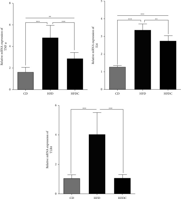
Methods: Rats were assigned into three groups each including ten rats: the control group (CD), the high-fat diet group (HFD), and the high-fat diet + curcumin (HFDC) group. Serum glucose, insulin, and triglycerides (TAGs) were observed. In addition, apoptosis (indicated by hepatic DNA fragmentation) and oxidative stress status (indicated by hepatic MPO, GSH, and SOD) were assessed. Histopathological examinations included the GIT (liver and duodenum) and heart in addition to quantitative real-time polymerase chain reaction (qRT-PCR) assays of the adipose tissue genetic expressions for inflammatory signaling pathways (TLR4, IL-6, and TNF-α).
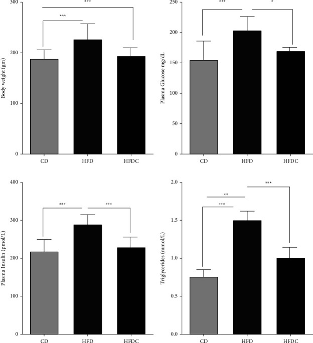
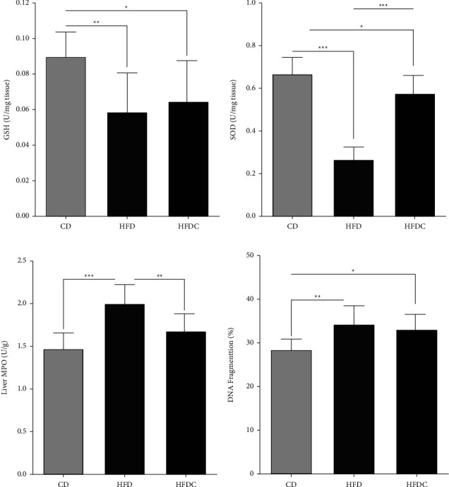
Results: The overall findings showed that the HFD group exhibited significantly higher levels of glucose, TAGs, and insulin than the control group (P < 0.01). The histological abnormalities of the studied organs in the HFD group were paralleled by these biochemical abnormalities, which were strongly associated with increased apoptosis, increased oxidative stress, and increased expression of the inflammatory signaling markers. There were significant improvements in the HFDC group in terms of biochemical, inflammatory, and histological investigations.
Conclusions: This study's findings concluded that obesity is significantly associated with biochemical and microscopic alterations in many organs. Curcumin exerted potent antitoxic, antioxidant, tissue-protective, and antiobesity effects. Curcumin is recommended to be added to various dietary regimens to prevent or delay the organs' dysfunction among obese people.
Copyright © 2022 Mohammed H. Hassan et al.
Conflict of interest statement
The authors declare that they have no conflicts of interest.
Figures
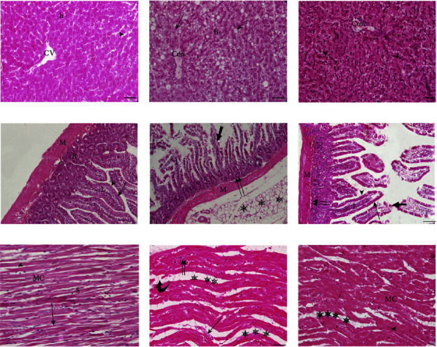
 ), and microvesicular steatosis (thin arrow). (d–f): Photomicrographs of male albino rat's duodenum stained with haematoxylin and eosin (HE). (×200 = 100 μm). (d): Duodenal photomicrograph of the CD group. (e): Duodenal photomicrograph of the HFD group. (f): Duodenal photomicrograph of the HFDC group. Musculosa (M), Brunner's glands (B), Lamina propria (L), simple columnar epithelium of villus (thin arrow), Goblet cells (arrow head), epithelial erosion (thick arrow), inflammatory infiltration (I), Necrosis (double arrow) of the inner circular muscle fibers of musculosa, Massive amounts of adipose tissues (stars). (g–i): Photomicrographs of male albino rat's heart stained with haematoxylin and eosin (HE). (×400 = 50 μm). (a): Heart photomicrograph of the CD group. (b): Heart photomicrograph of the HFD group. (c): Heart photomicrograph of the HFDC group. Muscle fibers (MC), the intercellular spaces (S), vesicular nuclei (arrow), intercalated discs (I) (arrow head), damage of cardiac muscle fibers (
 ), inflammatory cells (double arrows), separation of cardiac muscle fibers (stars), and extravasated red blood cells (thin arrow).
Similar articles
-
Preventive effect of curcumin on inflammation, oxidative stress and insulin resistance in high-fat fed obese rats.J Complement Integr Med. 2016 Jun 1;13(2):137-43. doi: 10.1515/jcim-2015-0070. J Complement Integr Med. 2016. PMID: 26845728
-
Dietary curcumin and capsaicin: Relationship with hepatic oxidative stress and apoptosis in rats fed a high fat diet.Adv Clin Exp Med. 2019 Aug;28(8):1013-1020. doi: 10.17219/acem/94145. Adv Clin Exp Med. 2019. PMID: 30993920
-
Trigonelline and curcumin alone, but not in combination, counteract oxidative stress and inflammation and increase glycation product detoxification in the liver and kidney of mice with high-fat diet-induced obesity.J Nutr Biochem. 2020 Feb;76:108303. doi: 10.1016/j.jnutbio.2019.108303. Epub 2019 Nov 23. J Nutr Biochem. 2020. PMID: 31812909
-
Evaluation of antiobesity and hepatorenal protective activities of Salvia officinalis extracts pre-treatment in high-fat diet-induced obese rats.Environ Sci Pollut Res Int. 2022 Oct;29(49):75043-75056. doi: 10.1007/s11356-022-21092-2. Epub 2022 Jun 1. Environ Sci Pollut Res Int. 2022. PMID: 35648345
-
Effect of dietary curcumin and capsaicin on testicular and hepatic oxidant-antioxidant status in rats fed a high-fat diet.Appl Physiol Nutr Metab. 2019 Jul;44(7):774-782. doi: 10.1139/apnm-2018-0622. Epub 2019 Jan 3. Appl Physiol Nutr Metab. 2019. PMID: 30605349
Cited by
-
Curcumin as a Potential Antioxidant in Stress Regulation of Terrestrial, Avian, and Aquatic Animals: A Review.Antioxidants (Basel). 2023 Aug 31;12(9):1700. doi: 10.3390/antiox12091700. Antioxidants (Basel). 2023. PMID: 37760003 Free PMC article. Review.
-
Nutrients Lowering Obesity-Linked Chemokines Blamable for Metastasis.Int J Mol Sci. 2025 Mar 4;26(5):2275. doi: 10.3390/ijms26052275. Int J Mol Sci. 2025. PMID: 40076892 Free PMC article. Review.
-
Dietary Curcumin Attenuates Hepatic Cellular Senescence by Suppressing the MAPK/NF-κB Signaling Pathway in Aged Mice.Antioxidants (Basel). 2023 May 27;12(6):1165. doi: 10.3390/antiox12061165. Antioxidants (Basel). 2023. PMID: 37371895 Free PMC article.
-
Adipo-Modulation by Turmeric Bioactive Phenolic Components: From Curcuma Plant to Effects.Int J Mol Sci. 2025 Jul 17;26(14):6880. doi: 10.3390/ijms26146880. Int J Mol Sci. 2025. PMID: 40725127 Free PMC article. Review.
-
Oral Treatment of Lactoferrin Nanocapsules Modulates the Immune Response of Mice to a Cryptosporidiosis Infection.Acta Parasitol. 2025 May 16;70(3):105. doi: 10.1007/s11686-025-01022-1. Acta Parasitol. 2025. PMID: 40377774 Free PMC article.
References
-
- Vehapoglu A., Turkmen S., Goknar N., Ozer O. F. Reduced antioxidant capacity and increased subclinical inflammation markers in prepubescent obese children and their relationship with nutritional markers and metabolic parameters. Redox Report . 2016;21(6):271–280. doi: 10.1080/13510002.2015.1133035. - DOI - PMC - PubMed
LinkOut - more resources
Full Text Sources
Research Materials
Miscellaneous
