Effects on Autophagy of Moxibustion at Governor Vessel Acupoints in APP/PS1double-Transgenic Alzheimer's Disease Mice through the lncRNA Six3os1/miR-511-3p/AKT3 Molecular Axis
- PMID: 36248429
- PMCID: PMC9556209
- DOI: 10.1155/2022/3881962
Effects on Autophagy of Moxibustion at Governor Vessel Acupoints in APP/PS1double-Transgenic Alzheimer's Disease Mice through the lncRNA Six3os1/miR-511-3p/AKT3 Molecular Axis
Abstract
Objective: To explore the effect and mechanism of moxibustion at acupoints of the governor vessel on lncRNA Six3os1 in amyloid precursor protein/presenilin1 (APP/PS1) double-transgenic Alzheimer's disease (AD) mice.
Methods: Twenty-four specific pathogen-free and APP/PS1 double-transgenic male mice were randomly allocated into the AD model and moxibustion groups, with 12 cases in each group. Twelve syngeneic C57BL/6J mice were selected as the control group. Mice in the moxibustion group received aconite cake-separated moxibustion at the Baihui acupoint. Suspension moxibustion was applied at Fengfu and Dazhui for 15 minutes each day. All treatments were conducted over two weeks. Control and AD model mice were routinely fed without any intervention. Behavioral observation tests were conducted before and after the intervention. The autophagosome in the hippocampus was observed using transmission electron microscopy. Immunohistochemistry was performed to detect Aβ1-42 expression. LC3B and P62 expressions were evaluated by immunofluorescence. The expression levels of the lncRNAs Six3os1, miR-511-3p, and AKT3 were detected by qRT-PCR. The differential expression of PI-3K, AKT3, mTOR, LC3B-II/I, and P62 proteins in the hippocampus was detected by western blot. The dual-luciferase assay was undertaken to examine the targeting relationships of the lncRNAs Six3os1, miR-511-3p, and AKT3.
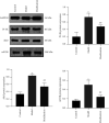
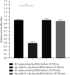
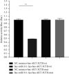
Results: Compared with the control group, the AD model showed higher escape latency in the Morris Water Maze and reduced autophagic vacuoles in the cytoplasm of hippocampal neurons (both p < 0.01). Compared with the control group, the AD model showed higher expression of Aβ1-42, the lncRNAs Six3os1, PI-3K, mTOR, P62, and AKT3 protein (all p < 0.01); but lower mir-511-3p and LC3B (both p < 0.01). Compared with the AD model group, the moxibustion group had a shorter escape latency, more autophagic bubbles in the hippocampus, and lower expression of positive Aβ1-42, the lncRNAs Six3os1, PI-3K, mTOR, P62, and AKT3 protein (all p < 0.01). In contrast, the levels of miR-511-3p and LC3B proteins were considerably increased in the moxibustion group compared to the AD model group (both p < 0.01). Based on the dual-luciferase assay, there was a targeting link among the lncRNAs Six3os1, miR-511-3p, and AKT3.
Conclusion: Moxibustion at acupoints of the governor vessel can suppress the lncRNA Six3os1 expression, promote cell autophagy, accelerate Aβ1-42 clearance and alleviate cognitive dysfunction of AD mediated by the PI3K/AKT/mTOR signaling pathway through the lncRNA Six3os1/miR-511-3p/AKT3 axis.
Copyright © 2022 Yu-Mei Jia et al.
Conflict of interest statement
The authors declare that they have no conflicts of interest.
Figures


Similar articles
-
[Effect of moxibustion on autophagy lysosome function mediated by mTOR/TFEB pathway and lncRNA H19 expression in APP/PS1 double transgenic mice].Zhen Ci Yan Jiu. 2022 Aug 25;47(8):665-72. doi: 10.13702/j.1000-0607.20211177. Zhen Ci Yan Jiu. 2022. PMID: 36036098 Chinese.
-
[Moxibustion at acpoints of governor vessel on regulating PI3K/Akt/mTOR signaling pathway and enhancing autophagy process in APP/PS1 double-transgenic Alzheimer's disease mice].Zhongguo Zhen Jiu. 2019 Dec 12;39(12):1313-8. doi: 10.13703/j.0255-2930.2019.12.015. Zhongguo Zhen Jiu. 2019. PMID: 31820607 Chinese.
-
[Moxibustion improves learning-memory ability by promoting cellular autophagy and regulating autophagy-related proteins in hippocampus and cerebral cortex in APP/PS1 transgenic Alzheimer's disease mice].Zhen Ci Yan Jiu. 2019 Apr 25;44(4):235-41. doi: 10.13702/j.1000-0607.180305. Zhen Ci Yan Jiu. 2019. PMID: 31056874 Chinese.
-
[Effect of moxibustion on autophagy in mice with Alzheimer's disease based on mTOR/p70S6K signaling pathway].Zhongguo Zhen Jiu. 2022 Sep 12;42(9):1011-6. doi: 10.13703/j.0255-2930.20210705-k0004. Zhongguo Zhen Jiu. 2022. PMID: 36075597 Chinese.
-
LncRNA BACE1-AS Promotes Autophagy-Mediated Neuronal Damage Through The miR-214-3p/ATG5 Signalling Axis In Alzheimer's Disease.Neuroscience. 2021 Feb 10;455:52-64. doi: 10.1016/j.neuroscience.2020.10.028. Epub 2020 Nov 13. Neuroscience. 2021. PMID: 33197504
Cited by
-
Ischemic Stroke and Autophagy: The Roles of Long Non-Coding RNAs.Curr Neuropharmacol. 2024;23(1):85-97. doi: 10.2174/1570159X22666240704123701. Curr Neuropharmacol. 2024. PMID: 39021183 Free PMC article. Review.
-
The Emerging Role of Autophagy-Associated lncRNAs in the Pathogenesis of Neurodegenerative Diseases.Int J Mol Sci. 2023 Jun 2;24(11):9686. doi: 10.3390/ijms24119686. Int J Mol Sci. 2023. PMID: 37298636 Free PMC article. Review.
-
lncRNA six3os1 diagnoses acute stroke, predicts disease severity, and predicts post-stroke cognitive impairment.BMC Neurol. 2024 Dec 26;24(1):491. doi: 10.1186/s12883-024-04003-5. BMC Neurol. 2024. PMID: 39722013 Free PMC article.
-
The Mechanism of Acupuncture Regulating Autophagy: Progress and Prospect.Biomolecules. 2025 Feb 11;15(2):263. doi: 10.3390/biom15020263. Biomolecules. 2025. PMID: 40001566 Free PMC article. Review.
References
LinkOut - more resources
Full Text Sources
Research Materials
Miscellaneous

