. 2022 Sep 30:13:995191.
doi: 10.3389/fimmu.2022.995191. eCollection 2022.

Pathogenic variant c.1052T>A (p.Leu351Gln) in adenosine deaminase 2 impairs secretion and elevates type I IFN responsive gene expression

Affiliations

Pathogenic variant c.1052T>A (p.Leu351Gln) in adenosine deaminase 2 impairs secretion and elevates type I IFN responsive gene expression

Sarah M Bowers et al. Front Immunol. .

Abstract

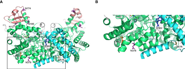
Background: Adenosine deaminase 2 (ADA2) is a homodimeric, extracellular enzyme and putative growth factor that is produced by cells of the myeloid lineage and, catalytically, deaminates extracellular adenosine to inosine. Loss-of-(catalytic)-function variants in the ADA2 gene are associated with Deficiency of ADA2 (DADA2), an autosomal recessive disease associated with an unusually broad range of inflammatory manifestations including vasculitis, hematological defects and cytopenia. Previous work by our group led to the identification of ADA2 variants of novel association with DADA2, among which was a unique c.1052T>A (p.Leu351Gln; herein referred to as L351Q) variant located in the catalytic domain of the protein.

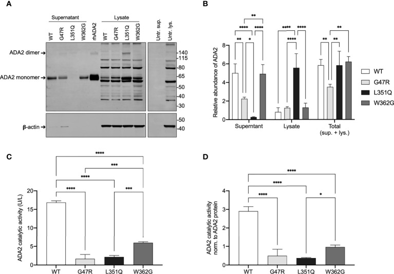
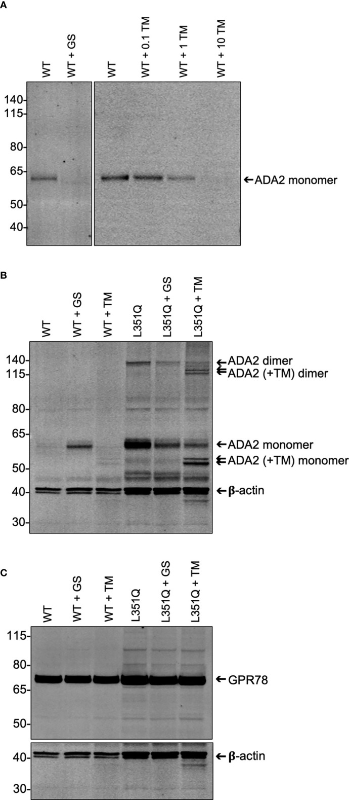
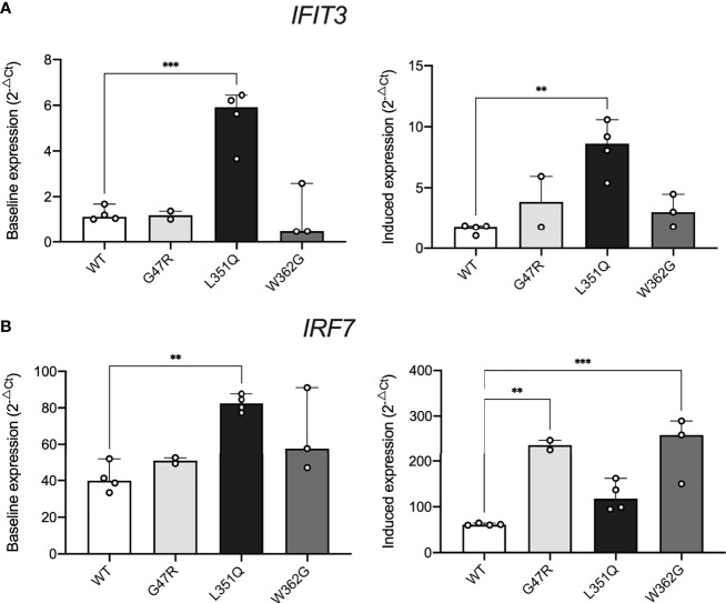
Methods: Mammalian (Flp-IN CHO) cells were engineered to stably express wild-type ADA2 and ADA2 protein variants, including the pathogenic L351Q variant identified in DADA2 patients. An enzyme assay and immunoblotting were used to assess ADA2 catalytic activity and secretion, respectively, and the outcome of experimentally induced inhibition of protein processing (Golgi transport and N-linked glycosylation) was assessed. Reverse transcription quantitative real-time PCR (RT-qPCR) was applied to determine the relative expression of Type I Interferon stimulated genes (ISGs), IFIT3 and IRF7.

Results: In addition to abrogating catalytic activity, the L351Q variant impaired secretion of L351Q ADA2 resulting in an intracellular accumulation of L351Q ADA2 protein that was not observed in cells expressing wild-type ADA2 or other ADA2 protein variants. Retention of L351Q ADA2 was not attributable to impaired glycosylation on neighboring asparagine residues and did not impact cell growth or integrity. Constitutive expression of Type I ISGs IFIT3 and IRF7 was observed in cells expressing L351Q ADA2.

Conclusions: The impaired secretion of L351Q ADA2 may be an important factor leading to the severe phenotype observed in patients with this variant further emphasizing the importance of assessing impacts beyond catalytic activity when evaluating genotype-phenotype relationships in DADA2.

Keywords: adenosine deaminase 2 (ADA2); inflammation; pediatric; systemic vasculitis; type I Interferon (IFN).


Conflict of interest statement

The authors declare that the research was conducted in the absence of any commercial or financial relationships that could be construed as a potential conflict of interest.

Figures

Figure 1
Figure 1
Predicted protein structure of human ADA2. (A) A ribbon diagram of the ADA2 homodimer crystallized in the presence of coformycin (transition state analog) and zinc (cofactor) using data available from the Protein Data Bank (PDB 3LGG) (16). The catalytic domain, dimerization domain, putative receptor binding (PRB) domain, and small unique elements are shown in green, cyan, light red, and grey, respectively. Predicted sites of N-linked glycosylation are labeled (N127, N174, N185, and N378) on one monomer, and the relevant asparagine residues are shown in purple (16, 17). Leucine 351 (L351) and tryptophan 362 [W362; W336 in (16)] are labelled and shown in orange and blue, respectively, on one monomer and glycine 47 (G47) is labelled and shown in yellow on the other monomer. The boxed region of A is expanded in (B). Figures were generated using PyMOL version 2.4.0 (Schrödinger, Inc., NY, USA).
Figure 2
Figure 2
ADA2 protein expression and catalytic activity in CHO cells transfected with expression vectors for wild-type and variant ADA2. (A) Representative anti-6xHis immunoblot (upper panel) and beta-actin (β-actin; loading control for lysates, lower panel) of supernatant and lysate (3x10⁵ cell equivalents) from Flp-IN CHO cells untransfected (Untr.) and transfected with expression vectors for His-tagged wild-type (WT) and variant (G47R, L351Q, W362G) adenosine deaminase 2 (ADA2) (n = 3). Recombinant human (rh) 6xHis-tagged rhADA2 (50 ng rhADA2) is included for comparison as is untransfected cell supernatant (Untr. sup.) and lysate (Untr. lys.). Ladder (left) marks relative molecular weight in kilodaltons (kDa). Arrows show ADA2 monomers (~60 kDa) and dimers (>120 kDa). (B) Graph of relative protein abundance (y-axis; Relative abundance of ADA2, n = 3) based on densitometry analysis of WT and variant ADA2 in cell supernatant (x-axis; Supernatant), lysate (x-axis; Lysate), and the calculated total from supernatant and lysate combined (x-axis; Total (sup. + lys.)), visualized by anti-6xHis and anti-β-actin immunoblots (as shown in A), with signal normalized to 50 ng of rhADA2 included on each gel (see Methods). (C) Total ADA2 catalytic activity (y-axis; ADA2 catalytic activity (U/L), n = 3) in supernatant and lysate from Flp-IN CHO cells transfected with expression vectors for WT (x-axis; WT, white) and variant (x-axis; G47R, light grey; L351Q, black; W362G, dark grey) ADA2. (D) Total ADA2 catalytic activity shown in (C), normalized to the total relative abundance of ADA2 protein (sup. + lys.) shown in (B) (y-axis; ADA2 catalytic activity norm. to ADA2 protein, n = 3). Bars show mean + SD and statistics show significant results of Tukey’s multiple comparisons test to compare group means. *p ≤ 0.05, **p ≤ 0.01, ***p ≤ 0.001, ****p≤ 0.0001.
Figure 3
Figure 3
Impact of experimentally-induced inhibition of protein processing on wildtype and L351Q ADA2 expression from Flp-IN CHO cells. (A) Representative (n = 3) anti-ADA2 immunoblot of supernatant from wild-type (WT) ADA2 expressing Flp-IN CHO cells treated without (WT), with Golgi transport inhibitor GolgiStop (WT + GS; 5.6 µL/mL) and with N-linked glycosylation inhibitor tunicamycin (WT + TM; 0 – 10 µg/mL). Arrow shows ADA2 monomers (~60 kDa). (B) Representative (n = 3) anti-ADA2 immunoblots of lysate (3 x 10⁵ cell equivalents) from Flp-IN CHO cells transfected with expression vectors for WT or L351Q ADA2 and treated with no inhibitor (WT, L351Q), GolgiStop (WT + GS, L351Q + GS; 5.6 µL/mL GolgiStop), or tunicamycin (WT + TM, L351Q + TM; 10 µg/mL tunicamycin). Samples were probed simultaneously for a loading control, beta-actin (β-actin). Arrows show ADA2 monomers and dimers in the absence and presence (+TM) of tunicamycin. (C) Representative (n = 3) anti-GPR78 immunoblots of lysate (3 x 10⁵ cell equivalents) from Flp-IN CHO cell samples shown in (B). For (A-C), ladder (left) marks relative molecular weights in kilodaltons (kDa).
Figure 4
Figure 4
Impact of intracellular L351Q ADA2 on cell stress. Expression of (A) IFIT3 and (B) IRF7 in Flp-IN CHO cells transfected with expression vectors for wild-type (WT, white) and G47R (light grey), L351Q (black), and W362G (dark grey) variant ADA2 cultured with no stimulus (left panels, y-axis; Baseline expression (2-ΔCt)) or recombinant human Type I IFNalpha 2b (10 ng/mL, 24 hr) (right panels, y-axis; Induced expression (2-ΔCt)). Gene expression was calculated using the delta Ct method (2-ΔCt), with eukaryotic HPRT1 as a housekeeping control; bars show mean + SD (n = 2-4). Statistics show significant results of Dunnett’s multiple comparisons test to compare group means to WT (WT = control). **p ≤ 0.01, ***p ≤ 0.001.

Similar articles

Cited by

References

    1. Navon Elkan P, Pierce SB, Segel R, Walsh T, Barash J, Padeh S, et al. . Mutant adenosine deaminase 2 in a polyarteritis nodosa vasculopathy. N Engl J Med (2014) 370:921–31. doi: 10.1056/NEJMoa1307362 - DOI - PubMed
    1. Zhou Q, Yang D, Ombrello AK, Zavialov AV, Toro C, Zavialov AV, et al. . Early-onset stroke and vasculopathy associated with mutations in ADA2. N Engl J Med (2014) 370:911–20. doi: 10.1056/NEJMoa1307361 - DOI - PMC - PubMed
    1. Gibson KM, Morishita KA, Dancey P, Moorehead P, Drögemöller B, Han X, et al. . Identification of novel adenosine deaminase 2 gene variants and varied clinical phenotype in pediatric vasculitis. Arthritis Rheumatol Hoboken NJ (2019) 71:1747–55. doi: 10.1002/art.40913 - DOI - PubMed
    1. Meyts I, Aksentijevich I. Deficiency of adenosine deaminase 2 (DADA2): Updates on the phenotype, genetics, pathogenesis, and treatment. J Clin Immunol (2018) 38:569–78. doi: 10.1007/s10875-018-0525-8 - DOI - PMC - PubMed
    1. Van Montfrans JM, Hartman EAR, Braun KPJ, Hennekam EAM, Hak EA, Nederkoorn PJ, et al. . Phenotypic variability in patients with ADA2 deficiency due to identical homozygous R169Q mutations. Rheumatol Oxf Engl (2016) 55:902–10. doi: 10.1093/rheumatology/kev439 - DOI - PubMed

Publication types