. 2022 Sep 29:13:931388.
doi: 10.3389/fimmu.2022.931388. eCollection 2022.

Sustained Infiltration of Neutrophils Into the CNS Results in Increased Demyelination in a Viral-Induced Model of Multiple Sclerosis

Affiliations

Sustained Infiltration of Neutrophils Into the CNS Results in Increased Demyelination in a Viral-Induced Model of Multiple Sclerosis

Dominic D Skinner et al. Front Immunol. .

Abstract

Intracranial inoculation of the neuroadapted JHM strain of mouse hepatitis virus (JHMV) into susceptible strains of mice results in acute encephalomyelitis followed by a cimmune-mediated demyelination similar to the human demyelinating disease multiple sclerosis (MS). JHMV infection of transgenic mice in which expression of the neutrophil chemoattractant chemokine CXCL1 is under the control of a tetracycline-inducible promoter active within GFAP-positive cells results in sustained neutrophil infiltration in the central nervous system (CNS) that correlates with an increase in spinal cord demyelination. We used single cell RNA sequencing (scRNAseq) and flow cytometry to characterize molecular and cellular changes within the CNS associated with increased demyelination in transgenic mice compared to control animals. These approaches revealed the presence of activated neutrophils as determined by expression of mRNA transcripts associated with neutrophil effector functions, including CD63, MMP9, S100a8, S100a9, and ASPRV1, as well as altered neutrophil morphology and protein expression. Collectively, these findings reveal insight into changes in the profile of neutrophils associated with increased white matter damage in mice persistently infected with a neurotropic coronavirus.

Keywords: chemokine receptors; chemokines; coronavirus; neuroinflammation; neutrophils.


Conflict of interest statement

The authors declare that the research was conducted in the absence of any commercial or financial relationships that could be construed as a potential conflict of interest.

Figures
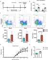
Figure 1
Figure 1
JHMV-infection of double-tg mice results in worsened clinical disease following doxycycline-induced CXCL1 expression. (A) Schematic overview showing experimental paradigm. Single-tg and double-tg mice were infected intracranially (i.c) with 250 PFU of JHMV and treated daily with doxycycline (50 mg/kg) via i.p. injection from 2-12 days p.i. Tissue was collected and analyzed at defined times. (B) ELISA was performed using supernatants of homogenized infected brain tissue at days 7 (n = 6-7 mice per group) and 12 (n = 4 mice per group) p.i.; double-tg mice showed elevated levels of CXCL1 protein at both timepoints following doxycycline treatment compared to single-tg control mice. Data is derived from a minimum of 2 independent experiments per timepoint. (C) Flow cytometry dot plots showing increased neutrophil (CD45+Ly6G+) accumulation in the blood, brain and spinal cords of double-tg mice compared to single-tg control. Data is derived from 2 independent experiments with a minimum of 5 mice used per tissue type. (D) Representative clinical disease showing JHMV-infected double-tg (n = 5) with increased (p < 0.05) clinical disease severity compared to infected single-tg mice (n = 6) at defined times p.i. (E) Viral titers are increased (p < 0.05) within the brains of JHMV-infected double-tg mice (n = 4) compared to infected single-tg mice (n = 4) at day 12 p.i. at day 12 p.i. *p< 0.05, **p< 0.01, ****p< 0.0001.
Figure 2
Figure 2
Demyelination is increased in doxycycline-treated double-tg infected with JHMV. (A) Representative H&E/LFB-stained spinal cord sections from doxycycline-treated double-tg and single-tg mice infected with JHMV at day 14 p.i. reveals increased spinal cord demyelination (red dashed line) in double-tg mice compared to single-tg mice. (B) Quantification of demyelination severity indicates white matter damage is significantly (p < 0.01) more severe in infected double-tg mice (n = 5) compared to single tg mice (n = 6); data is derived from 2 independent experiments. **p< 0.01. (C) Flow cytometric analysis of neutrophils present in the spinal cords of uninfected doxycycline-treated single-tg mice (n = 3) and double-tg mice (n = 3) shows no significant difference in neutrophil numbers between experimental groups. (D) Representative H&E/LFB-stained spinal cord sections from uninfected doxycycline-treated single-tg mice (n = 4) and double-tg (n = 4) reveals no evidence of demyelination.
Figure 3
Figure 3
scRNAseq of CD45+ cells isolated from spinal cords of JHMV-infected mice.(A) Uniform Manifold Approximation and Projection (UMAP) plot of scRNASeq data revealing 14 distinct cell clusters (aggregate data from JHMV-infected double-tg (n = 4) and single-tg mice (n = 4) treated with doxycycline at 12 days p.i. (B) Heat map showing the top 5 differentially expressed genes with each cluster. Columns show each cluster, with sub columns representing single-tg and double-tg mice; rows specify genes. (C) UMAP plot displaying an overlay of single-tg (blue) and double-tg (red) scRNASeq data. (D) Number of cells per cluster comparing single-tg (blue) and double-tg (red) scRNASeq data from spinal cord at 12 days p.i.
Figure 4
Figure 4
Neutrophils isolated from the spinal cord express genes associated with chemotaxis and demyelination. (A) UMAP plot showing highlighted (green) neutrophil cluster from JHMV-infected single-tg (n = 4) and double-tg (n = 4) mice. (B–H) Violin plots show expression of Asprv1, Cd63, Cxcr2 Lcn2 (NGAL), Mmp-9, S100a8, S100a9 primarily within neutrophil clusters; black dots represent individual cells. Inset UMAP plots show the expression level of each gene within the neutrophil cluster. Bar graphs show the number of positive neutrophils for each respective gene for double-tg (red) and single-tg (blue) datasets. (I) Violin plots demonstrating that neutrophils from both JHMV-infected single-tg and double-tg mice express Asprv1, Cd63, Cxcr2, Lcn2 (NGAL), Mmp-9, S100a8, and S100a9 transcripts. (J) Pie charts show the frequency of Cxcr2+ neutrophils from the double-tg mice that express Asprv1, Cd63, Lcn2 (NGAL), Mmp-9, S100a8, S100a9.
Figure 5
Figure 5
Neutrophil accumulation alters expression of T cell attractant chemokines and T cell activation state. (A) Split violin plot of scRNAseq data showing Cxcr3 expression level (y-axis) compared by cluster (x-axis) with width of plot signifying frequency of Cxcr3 expressing cells. Cxcr3 expression relegated to T cell cluster subsets (CD4, CD8 1, CD8 2, Cyc. T Cells) in both single-tg (blue) and double-tg (red) mouse datasets. (B) Expression levels of chemokines Cxcl10 and Cxcl9 in monocytes are shown for single-tg (blue) and double-tg (red) mouse datasets. (C) Gene set enrichment analysis (GSEA) for IFN-γ responses in CD8 1 T cell and CD8 2 T cell clusters from spinal cord of single-tg (n = 4) and double-tg mice (n = 4) at day12 p.i. Responses to IFN-γ were enriched in both populations of double-tg CD8 T cells compared to control single-tg mice. (D) GSEA plot for inflammatory response and IFN-γ response in CD4 T cells. Responses to both inflammatory response and IFN-γ response was enriched in the CD4 T cell population of double-tg mice compared to control single-tg mice. The GSEA plots correspond to the designated Hallmark Inflammatory Response and Hallmark Interferon Gamma Response gene sets established in the Molecular Signatures Database (MSigDB) (45, 46). ***p < 0.001; ****p < 0.0001.
Figure 6
Figure 6
Neutrophils from JHMV-infected double-tg mice exhibit increased activation state (A) Representative flow plots reveal CD63+ neutrophils in the blood, brain, and spinal cord of JHMV-infected double-tg mice (n = 5) compared to infected single-tg (n = 7) control at day 12 p.i. Numbers of neutrophils in the spinal cord of infected double-tg mice are significantly (p < 0.05) increased compared to infected single-tg mice. (B) Representative flow plots showing mature neutrophils (CD45+Ly6g+CXCR4hiCD62Llow) in blood, brain and spinal cords of JHMV-infected double-tg mice (n = 5) and infected single-tg mice (n = 7). (C) Representative ImageStream flow cytometry on neutrophils (CD45+Ly6G+) isolated from blood, brain and spinal cord obtained of JHMV-infected double-tg mice reveals enhanced CD63 expression in spinal cord-associated neutrophils. In addition, neutrophils from brains and spinal cords exhibited increased granularity compared to neutrophils obtained from the blood. For data in panels (A–C), n = 5-7 mice per tissue type per group; 2 experiments. Representative flow plots are shown in panels (A, B). (D) JHMV-infected double-tg and single-tg mice were sacrificed at days 7 (n = 6-7 mice per group) and 12 p.i. (n = 4 mice per group) and levels of Neutrophil Elastase, NGAL, S100a8, and S100a9 determined by ELISA (3 independent experiments). *p< 0.05, **p< 0.01.

Similar articles

Cited by

References

    1. Lane TE, Hosking MP. The pathogenesis of murine coronavirus infection of the central nervous system. Crit Rev Immunol (2010) 30(2):119–30. doi: 10.1615/critrevimmunol.v30.i2.20 - DOI - PMC - PubMed
    1. Lane TE, Buchmeier MJ. Murine coronavirus infection: a paradigm for virus-induced demyelinating disease. Trends Microbiol (1997) 5(1):9–14. doi: 10.1016/S0966-842X(97)81768-4 - DOI - PMC - PubMed
    1. Bergmann CC, Lane TE, Stohlman SA. Coronavirus infection of the central nervous system: host-virus stand-off. Nat Rev Microbiol (2006) 4(2):121–32. doi: 10.1038/nrmicro1343 - DOI - PMC - PubMed
    1. Bender SJ, Weiss SR. Pathogenesis of murine coronavirus in the central nervous system. J Neuroimmune Pharmacol (2010) 5(3):336–54. doi: 10.1007/s11481-010-9202-2 - DOI - PMC - PubMed
    1. Hosking MP, Lane TE. The role of chemokines during viral infection of the CNS. PloS Pathog (2010) 6(7):e1000937–e1000937. doi: 10.1371/journal.ppat.1000937 - DOI - PMC - PubMed

Publication types