. 2022 Sep 30:13:1012647.
doi: 10.3389/fimmu.2022.1012647. eCollection 2022.

Soluble CD83 improves and accelerates wound healing by the induction of pro-resolving macrophages

Affiliations

Soluble CD83 improves and accelerates wound healing by the induction of pro-resolving macrophages

Dmytro Royzman et al. Front Immunol. .

Abstract

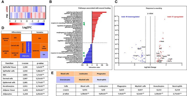
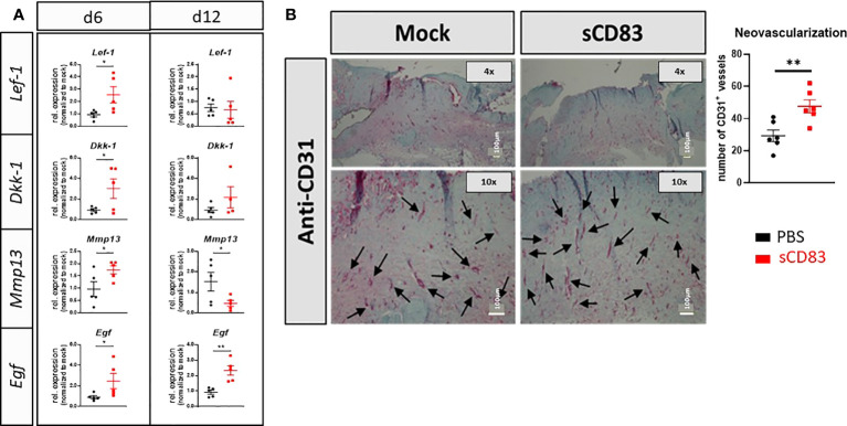
To facilitate the recovery process of chronic and hard-to-heal wounds novel pro-resolving treatment options are urgently needed. We investigated the pro-regenerative properties of soluble CD83 (sCD83) on cutaneous wound healing, where sCD83 accelerated wound healing not only after systemic but also after topical application, which is of high therapeutic interest. Cytokine profile analyses revealed an initial upregulation of inflammatory mediators such as TNFα and IL-1β, followed by a switch towards pro-resolving factors, including YM-1 and IL-10, both expressed by tissue repair macrophages. These cells are known to mediate resolution of inflammation and stimulate wound healing processes by secretion of growth factors such as epidermal growth factor (EGF) and vascular endothelial growth factor (VEGF), which promote vascularization as well as fibroblast and keratinocyte differentiation. In conclusion, we have found strong wound healing capacities of sCD83 beyond the previously described role in transplantation and autoimmunity. This makes sCD83 a promising candidate for the treatment of chronic- and hard-to-heal wounds.

Keywords: CD83; macrophages; stem cells; therapy; tissue regeneration; wound healing.


Conflict of interest statement

The authors declare that the research was conducted in the absence of any commercial or financial relationships that could be construed as a potential conflict of interest.

Figures

Figure 1
Figure 1
sCD83-treatment accelerates wound healing and modulates cellular composition within the wound beds. Wounds were inflicted in anesthetized 7-weeks-old female C57BL/6 mice using a 6 mm biopsy punch. Administration of 100 µg sCD83 was performed systemically until day 7 on a daily basis. (A) Representative images of wound areas on day 0, 3, 6, 7, and 12. (B) Wound size was determined using caliper and expressed as percentage of wound closure relatively to the day 0 value (each group and time point n≥12). (C) Representative H&E slides of day 7 wound biopsies. (D) Cellular composition within the wound biopsies of sCD83- and mock-treated mice, as assessed by flow cytometric analyses of day 6 samples (n=3). (E) Representative MELC-images (900µm x 900µm) of day 6 biopsy samples from PBS- and sCD83-treated mice [(E); left-hand side] along with the corresponding quantification of signal intensity as determined using ImageJ with n=3 [(E); right-hand side]. Dashed lines mark the wound site. Data are illustrated as mean ± SEM. Statistical analyses: (B) Two way ANOVA. Asterisks mark statistically significant difference (*p < 0.05 and **p < 0.01). The absence of asterisks indicates that there is no statistical significance.
Figure 2
Figure 2
sCD83-treatment changes the transcriptome profile towards a wound healing phenotype. RNA was extracted from day 3 wound biopsies and used for RNA sequencing analyses. The transcriptome of sCD83-treated mice was compared to mock controls. (A) Representative heat map of differently expressed transcripts in wound biopsies of sCD83-treated and PBS-treated mice on day 3: induction = red and suppression = blue (each n=2). (B) Pathway analysis was performed based on the transcriptome analyses and its interaction value with the corresponding pathways (red = induced and blue = suppressed). Green triangles highlight two wound healing-associated pathways that were induced or suppressed by sCD83, respectively. (C) Volcano plot of RNA sequencing analyses of wound biopsies of sCD83- vs. PBS-treated mice on day 3 included in the response to wounding pathway. Dots depicted on the right-hand side of the logFold change 0 value represent significantly upregulated gene transcripts (red), while the dots on the left-hand side represent significantly downregulated transcripts (blue) with logFC ≥ 0.585, respectively. The Y-axis depicts the p value. (D) Transcriptome data were further analysed using the Ingenuity Pathway Analysis (IPA) software from QIAGEN’s revealing enhanced wound healing pathways in sCD83-treated mice. (E) Cellular composition within day 3 wound sites of sCD83-treated mice as assessed from the RNA sequencing data by IPA.
Figure 3
Figure 3
sCD83 promotes MΦ differentiation and activity in the course of wound healing. (A) Transcript analyses were performed with skin biopsies relatively to the day 0 value (each group and time point n≥4). (B) RNA induction levels of Tnfα, Il-10, Msr-1 and Vegf were assessed in sCD83-treated MΦ under steady state conditions or in the presence of IL-4 (n=3) with the control group shown in black and sCD83-treated mice in red. (C) Ex vivo generated MΦ, cultured in the presence or absence of sCD83, were adoptively transferred into the wound beds and wound closure was assessed by caliper on day 3 relatively to the day 0 value (each n=5). Red circles highlight the wound area. (A) Two-way ANOVA. (B) Mann-Whitney test and (C) One-way ANOVA with F= 11,03. Asterisks mark statistically significant difference (* p< 0.05, **p < 0.01 and ***p < 0.001). The absence of asterisks indicates that there is no statistical significance.
Figure 4
Figure 4
sCD83 promotes epithelial stem cell activity during wound healing processes. (A) RT-PCR analyses were performed on day 6 and day 12 using wound biopsies from sCD83- or mock-treated mice with n≥5. (B) Blood vessel formation was assessed by immunohistochemical stainings, using a CD31-specific antibody. Vessel are highlighted by arrows along with the graphical representation of CD31+ vessels right-hand side; n=6). Data are illustrated as mean ± SEM. Statistical analyses were performed using Mann-Whitney test. Asterisks mark statistically significant difference (*p< 0.05 and **p< 0.01). The absence of asterisks indicates that there is no statistical significance.
Figure 5
Figure 5
Topical sCD83 application induces local wound healing. Wound lesions were induced using 6 mm punches, and diameters were measured by caliper, at indicated time points. (A) Western blot analyses of sCD83-hydrogel matrices after 10 days at 4°C, demonstrate the stability of the sCD83 protein. (B) Daily topical administration of sCD83 on days 1, 3 and 5 significantly increased wound healing. (C) Percentage of wound closure was set relatively to the day 0 value, with each group n=10. (D) Representative histological analyses of day 7 samples. Dashed lines mark the former wound sites. Data are illustrated as mean ± SEM and statistically analysed using One-way ANOVA for C. Asterisks mark statistically significant difference (*p < 0.05, ***p < 0.001 and ****p < 0.0001). The absence of asterisks indicates that there is no statistical significance.
Figure 6
Figure 6
Graphical Overview: sCD83-treatment accelerates cutaneous wound healing modulating the three phases of wound closure. sCD83 boosts the natural kinetics of the inflammatory-, proliferative- and remodeling phases, during the wound healing process. Initially, the inflammatory phase, within wound sites of sCD83-treated mice, was increased and prolonged, as indicated by upregulated TNFα and IL-1β levels, while IL-10-expression was delayed, until the proliferative phase. During wound healing YM-1+ sCD83 induced resolving MΦ are responsible for the secretion of IL-10 and the resolution of inflammation. Concomitantly, growth factors such as EGF and MMP remodeling enzymes are released by alternatively activated MΦ in order to restore tissue function and quality. Furthermore, the canonical Wnt-signaling cascade, which is related to epithelial stem cell proliferation, is boosted at earlier time points in order to replace the ablated tissue. The biological consequences are reflected in the increased presence of blood vessels in wounds of sCD83-treated mice.

Similar articles

Cited by

References

    1. Eming SA, Martin P, Tomic-Canic M. Wound repair and regeneration: Mechanisms, signaling, and translation. Sci Transl Med (2014) 6(265):265sr6. doi: 10.1126/scitranslmed.3009337 - DOI - PMC - PubMed
    1. Zarbock A, Polanowska-Grabowska RK, Ley K. Platelet-neutrophil-interactions: Linking hemostasis and inflammation. Blood Rev (2007) 21(2):99–111. doi: 10.1016/j.blre.2006.06.001 - DOI - PubMed
    1. Daley JM, Reichner JS, Mahoney EJ, Manfield L, Henry WL, Jr., Mastrofrancesco B, et al. . Modulation of macrophage phenotype by soluble product(s) released from neutrophils. J Immunol (2005) 174(4):2265–72. doi: 10.4049/jimmunol.174.4.2265 - DOI - PubMed
    1. Mosser DM, Edwards JP. Exploring the full spectrum of macrophage activation. Nat Rev Immunol (2008) 8(12):958–69. doi: 10.1038/nri2448 - DOI - PMC - PubMed
    1. Li Z, Ju X, Silveira PA, Abadir E, Hsu WH, Hart DNJ, et al. . CD83: Activation marker for antigen presenting cells and its therapeutic potential. Front Immunol (2019) 10:1312. doi: 10.3389/fimmu.2019.01312 - DOI - PMC - PubMed

Publication types

MeSH terms

Substances