

Neutrophil depletion in the pre-implantation phase impairs pregnancy index, placenta and fetus development
- PMID: 36248911
- PMCID: PMC9558710
- DOI: 10.3389/fimmu.2022.969336
Neutrophil depletion in the pre-implantation phase impairs pregnancy index, placenta and fetus development
Abstract
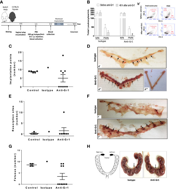
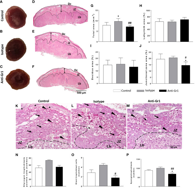
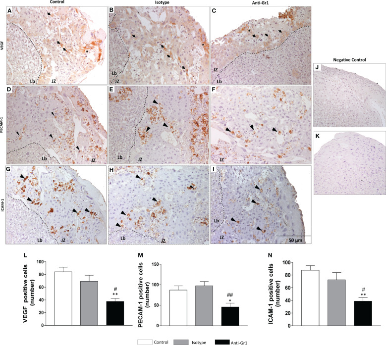
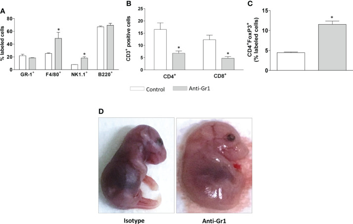
Maternal neutrophils cells are players in gestational tolerance and fetus delivery. Nonetheless, their actions in each phase of the pregnancy are unknown. We here investigated the role of maternal neutrophil depletion before the blastocyst implantation phase and outcomes in the pregnancy index, placenta, and fetus development. Neutrophils were pharmacologically depleted by i.p. injection of anti-Gr1 (anti-neutrophils; 200 µg) 24 hours after plug visualization in allogeneic-mated C57BL/6/BALB/c mice. Depletion of peripheral neutrophils lasted until 48 hours after anti-Gr1 injection (gestational day 1.5-3.5). On gestational day 5.5, neutrophil depletion impaired the blastocyst implantation, as 50% of pregnant mice presented reduced implantation sites. On gestational day 18.5, neutrophil depletion reduced the pregnancy rate and index, altered the placenta disposition in the uterine horns, and modified the structure of the placenta, detected by reduced junctional zone, associated with decreased numbers of giant trophoblast cells, spongiotrophoblast. Reduced number of placenta cells labeled for vascular endothelial growth factor (VEGF), platelet-endothelial cell adhesion molecule (PECAM-1), and intercellular cell adhesion molecule (ICAM-1), important markers of angiogenesis and adhesiveness, were detected in neutrophil depleted mice. Furthermore, neutrophil depletion promoted a higher frequency of monocytes, natural killers, and T regulatory cells, and lower frequency of cytotoxic T cells in the blood, and abnormal development of offspring. Associated data obtained herein highlight the pivotal role of neutrophils actions in the early stages of pregnancy, and address further investigations on the imbricating signaling evoked by neutrophils in the trophoblastic interaction with uterine epithelium.
Keywords: abnormal fetal development; anti-granulocytes; maternal immune system; placenta morphology; pregnancy index; window of implantation.
Copyright © 2022 Hebeda, Savioli, Scharf, de Paula-Silva, Gil, Farsky and Sandri.
Conflict of interest statement
The authors declare that the research was conducted in the absence of any commercial or financial relationships that could be construed as a potential conflict of interest.
Figures
Similar articles
-
Significance of platelet endothelial cell adhesion molecule-1 (PECAM-1) and intercellular adhesion molecule-1 (ICAM-1) expressions in preeclamptic placentae.Endocrine. 2012 Aug;42(1):125-31. doi: 10.1007/s12020-012-9644-9. Epub 2012 Mar 7. Endocrine. 2012. PMID: 22396143
-
Neutrophils induce proangiogenic T cells with a regulatory phenotype in pregnancy.Proc Natl Acad Sci U S A. 2016 Dec 27;113(52):E8415-E8424. doi: 10.1073/pnas.1611944114. Epub 2016 Dec 12. Proc Natl Acad Sci U S A. 2016. PMID: 27956610 Free PMC article.
-
ICAM-1-activated Src and eNOS signaling increase endothelial cell surface PECAM-1 adhesivity and neutrophil transmigration.Blood. 2012 Aug 30;120(9):1942-52. doi: 10.1182/blood-2011-12-397430. Epub 2012 Jul 17. Blood. 2012. PMID: 22806890 Free PMC article.
-
Melatonin and stable circadian rhythms optimize maternal, placental and fetal physiology.Hum Reprod Update. 2014 Mar-Apr;20(2):293-307. doi: 10.1093/humupd/dmt054. Epub 2013 Oct 16. Hum Reprod Update. 2014. PMID: 24132226 Review.
-
An integrated model of preeclampsia: a multifaceted syndrome of the maternal cardiovascular-placental-fetal array.Am J Obstet Gynecol. 2022 Feb;226(2S):S963-S972. doi: 10.1016/j.ajog.2020.10.023. Epub 2021 Mar 9. Am J Obstet Gynecol. 2022. PMID: 33712272 Review.
Cited by
-
Immune dysfunction mediated by the competitive endogenous RNA network in fetal side placental tissue of polycystic ovary syndrome.PLoS One. 2024 Mar 21;19(3):e0300461. doi: 10.1371/journal.pone.0300461. eCollection 2024. PLoS One. 2024. PMID: 38512862 Free PMC article.
-
Generation and characterization of a mouse model of conditional Chd4 knockout in the endometrial epithelium.bioRxiv [Preprint]. 2025 Jun 8:2025.06.05.658183. doi: 10.1101/2025.06.05.658183. bioRxiv. 2025. PMID: 40501592 Free PMC article. Preprint.
-
Exploring the Immunological Aspects and Treatments of Recurrent Pregnancy Loss and Recurrent Implantation Failure.Int J Mol Sci. 2025 Feb 3;26(3):1295. doi: 10.3390/ijms26031295. Int J Mol Sci. 2025. PMID: 39941063 Free PMC article. Review.
References
Publication types
MeSH terms
Substances
LinkOut - more resources
Full Text Sources
Miscellaneous