

RHOA G17V induces T follicular helper cell specification and involves angioimmunoblastic T-cell lymphoma via upregulating the expression of PON2 through an NF-κB-dependent mechanism
- PMID: 36249275
- PMCID: PMC9559328
- DOI: 10.1080/2162402X.2022.2134536
RHOA G17V induces T follicular helper cell specification and involves angioimmunoblastic T-cell lymphoma via upregulating the expression of PON2 through an NF-κB-dependent mechanism
Abstract
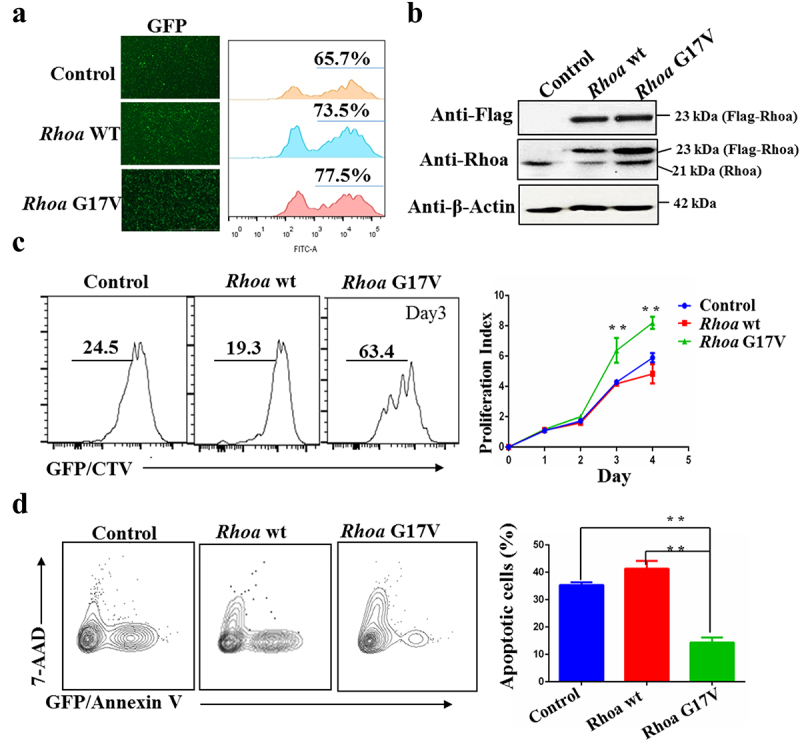
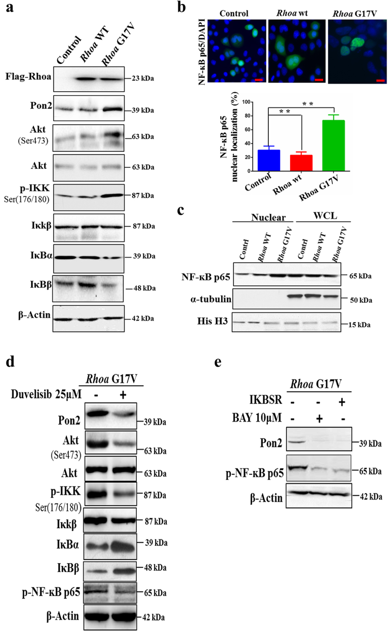
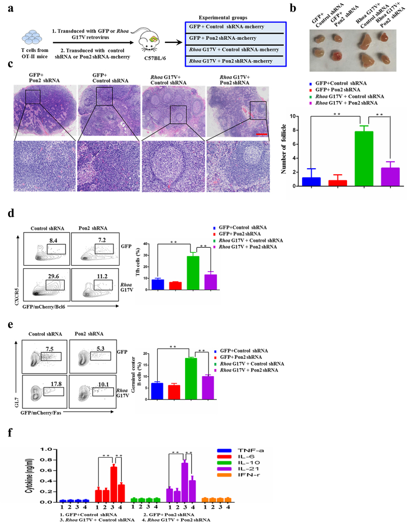
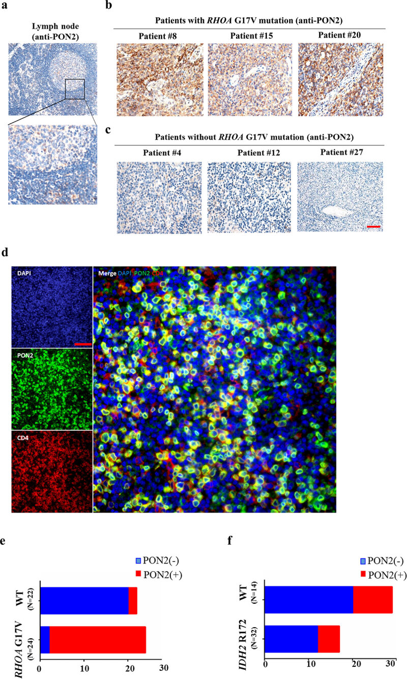
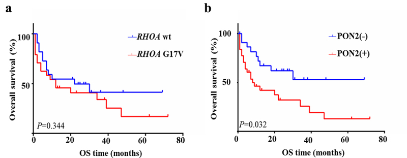
Angioimmunoblastic T-cell lymphoma (AITL) is a malignant hematologic tumor arising from T follicular helper (Tfh) cells. High-throughput genomic sequencing studies have shown that AITL is characterized by a novel highly recurring somatic mutation in RHOA, encoding p.Gly17Val (RHOA G17V). However, the specific role of RHOA G17V in AITL remains unknown. Here, we demonstrated that expression of Rhoa G17V in CD4+ T cells increased cell proliferation and induces Tfh cell specification associated with Pon2 upregulation through an NF-κB-dependent mechanism. Further, loss of Pon2 attenuated oncogenic function induced by genetic lesions in Rhoa. In addition, an abnormality of RHOA G17V mutation and PON2 expression is also detected in patients with AITL. Our findings suggest that PON2 associated with RHOA G17V mutation might control the direction of the molecular agents-based AITL and provide a new therapeutic target in AITL.
Keywords: AITL; NF-κB pathway; PON2; RHOA; mutation.
© 2022 The Author(s). Published with license by Taylor & Francis Group, LLC.
Conflict of interest statement
No potential conflicts of interest are disclosed.
Figures

Similar articles
-
RHOA G17V Induces T Follicular Helper Cell Specification and Promotes Lymphomagenesis.Cancer Cell. 2018 Feb 12;33(2):259-273.e7. doi: 10.1016/j.ccell.2018.01.001. Epub 2018 Feb 2. Cancer Cell. 2018. PMID: 29398449 Free PMC article.
-
Clinicopathologic Analysis of Angioimmunoblastic T-cell Lymphoma With or Without RHOA G17V Mutation Using Formalin-fixed Paraffin-embedded Sections.Am J Surg Pathol. 2016 Aug;40(8):1041-50. doi: 10.1097/PAS.0000000000000651. Am J Surg Pathol. 2016. PMID: 27158755
-
Angioimmunoblastic T-cell Lymphomas With the RHOA p.Gly17Val Mutation Have Classic Clinical and Pathologic Features.Am J Surg Pathol. 2016 Mar;40(3):335-41. doi: 10.1097/PAS.0000000000000555. Am J Surg Pathol. 2016. PMID: 26574844
-
Clinicopathological Implications of RHOA Mutations in Angioimmunoblastic T-Cell Lymphoma: A Meta-analysis: RHOA mutations in AITL.Clin Lymphoma Myeloma Leuk. 2021 Jul;21(7):431-438. doi: 10.1016/j.clml.2021.03.002. Epub 2021 Mar 19. Clin Lymphoma Myeloma Leuk. 2021. PMID: 33849798
-
G17V RHOA: Genetic evidence of GTP-unbound RHOA playing a role in tumorigenesis in T cells.Small GTPases. 2015;6(2):100-3. doi: 10.4161/21541248.2014.988088. Epub 2015 Jun 23. Small GTPases. 2015. PMID: 26103434 Free PMC article. Review.
Cited by
-
Binding of RHOA G17V to p300 enhances its HAT activity: a new mechanism of epigenetic deregulation in TFH lymphoma.Blood Adv. 2025 Jun 24;9(12):3031-3043. doi: 10.1182/bloodadvances.2024014671. Blood Adv. 2025. PMID: 40020168 Free PMC article.
-
Ten-Eleven Translocation Family Proteins: Structure, Biological Functions, Diseases, and Targeted Therapy.MedComm (2020). 2025 Jul 1;6(7):e70245. doi: 10.1002/mco2.70245. eCollection 2025 Jul. MedComm (2020). 2025. PMID: 40599235 Free PMC article. Review.
-
Advances in the pathogenesis and therapeutic strategies of angioimmunoblastic T-cell lymphoma.Clin Exp Med. 2023 Dec;23(8):4219-4235. doi: 10.1007/s10238-023-01197-9. Epub 2023 Sep 27. Clin Exp Med. 2023. PMID: 37759042 Review.
-
Genome-Wide Analysis of Sheep Artificially or Naturally Infected with Gastrointestinal Nematodes.Genes (Basel). 2023 Jun 26;14(7):1342. doi: 10.3390/genes14071342. Genes (Basel). 2023. PMID: 37510248 Free PMC article.
-
Prognostic prediction of lung adenocarcinoma by integrative analysis of RHOH expression and methylation.Clin Respir J. 2023 Mar;17(3):148-156. doi: 10.1111/crj.13574. Epub 2023 Jan 29. Clin Respir J. 2023. PMID: 36710485 Free PMC article.
References
-
- Dupuis J, Boye K, Martin N, Copie-Bergman C, Plonquet A, Fabiani B, Baglin A-C, Haioun C, Delfau-Larue M-H, Gaulard P, et al. Expression of cxcl13 by neoplastic cells in angioimmunoblastic t-cell lymphoma (aitl): a new diagnostic marker providing evidence that aitl derives from follicular helper t cells. Am J Surg Pathol. 2006;30(4):490–494. doi:10.1097/00000478-200604000-00009. - DOI - PubMed
-
- Grogg KL, Attygalle AD, Macon WR, Remstein ED, Kurtin PJ, Dogan A. Expression of cxcl13, a chemokine highly upregulated in germinal center t-helper cells, distinguishes angioimmunoblastic t-cell lymphoma from peripheral t-cell lymphoma, unspecified. Mod pathol. 2006;19(8):1101–1107. doi:10.1038/modpathol.3800625. - DOI - PubMed
Publication types
MeSH terms
Substances
LinkOut - more resources
Full Text Sources
Research Materials