. 2021 May 25;1(2):100028.
doi: 10.1016/j.xops.2021.100028. eCollection 2021 Jun.

PEX6 Mutations in Peroxisomal Biogenesis Disorders: An Usher Syndrome Mimic

Affiliations

PEX6 Mutations in Peroxisomal Biogenesis Disorders: An Usher Syndrome Mimic

Matthew D Benson et al. Ophthalmol Sci. .

Abstract

Purpose: Peroxisomal biogenesis disorders (PBDs) represent a spectrum of conditions that result in vision loss, sensorineural hearing loss, neurologic dysfunction, and other abnormalities resulting from aberrant peroxisomal function caused by mutations in PEX genes. With no treatments currently available, we sought to investigate the disease mechanism in a patient with a PBD caused by defects in PEX6 and to probe whether overexpression of PEX6 could restore peroxisome function and potentially offer therapeutic benefit.

Design: Laboratory-based study.

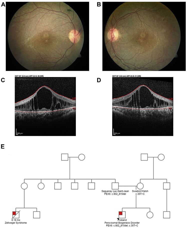
Participants: A 12-year-old boy sought treatment with hearing loss and retinopathy. After negative results in an Usher syndrome panel, targeted genetic testing revealed compound heterozygous mutations in PEX6. These included a 14-nucleotide deletion (c.802_815del: p.(Asp268Cysfs∗8)) and a milder missense variant (c.35T→C:(p.Phe12Ser)).

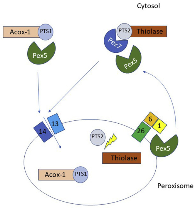
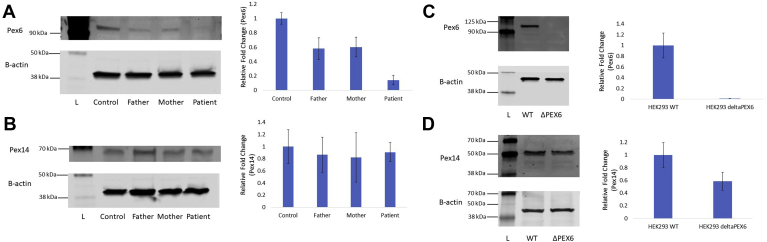
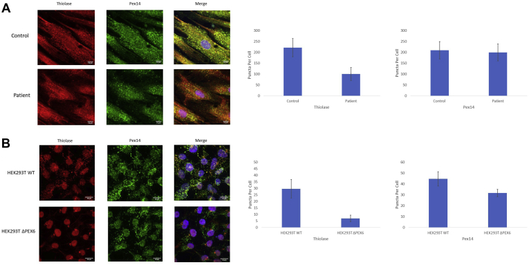
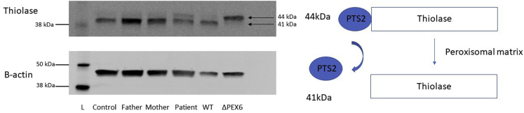
Methods: Patient-derived skin fibroblasts were cultured, and a PEX6 knockout cell line was developed using clustered regularly interspaced short palindromic repeats and Cas9 technology in HEK293T cells to emulate a more severe disease phenotype. Immunoblot analysis of whole cell lysates was performed to assess peroxisome number. Immunofluorescence studies used antibodies against components of the peroxisomal protein import pathway to interrogate the effects of mutations in PEX6 on protein trafficking.

Main outcome measures: Primary outcome measures were peroxisome abundance and matrix protein import.

Results: Peroxisome number was not significantly different between control fibroblasts and patient fibroblasts; however, fewer peroxisomes were observed in PEX6 knockout cells compared with wild-type cells (P = 0.04). Analysis by immunofluorescent microscopy showed significantly impaired peroxisomal targeting signal 1- and peroxisomal targeting signal 2-mediated matrix protein import in both patient fibroblasts and PEX6 knockout cells. Overexpressing PEX6 resulted in improved matrix protein import in PEX6 knockout cells.

Conclusions: Mutations in PEX6 were responsible for combined hearing loss and retinopathy in our patient. The primary peroxisomal defect in our patient's skin fibroblasts was impaired peroxisomal protein import as opposed to reduction in the number of peroxisomes. Genetic strategies that introduce wild-type PEX6 into cells deficient in PEX6 protein show promise in restoring peroxisome function. Future studies of patient-specific induced pluripotent stem cell-derived retinal pigment epithelium cells may clarify the role of PEX6 in the retina and the potential for gene therapy in these patients.

Keywords: CRISPR, clustered regularly interspaced short palindromic repeats; DTM, docking translocation module; GFP, green fluorescent protein; HEK293T, human embryonic kidney 293T; Hearing loss; PBD, peroxisomal biogenesis disorder; PBS, phosphate-buffered saline; PEX6; PTS1, peroxisomal targeting signal 1; PTS2, peroxisomal targeting signal 2; Peroxisomal biogenesis disorders; Peroxisome; RPE, retinal pigment epithelium; Retinal degeneration; Usher syndrome; WT, wild-type.


Figures

Figure 1
Figure 1
Illustration of a model of peroxisomal matrix protein import. Nascent polypeptides synthesized in the cytosol, destined for the peroxisomal lumen, are labeled with either a C-terminal peroxisomal targeting signal 1 (PTS1) tripeptide or an N-terminal peroxisomal targeting signal 2 (PTS2) nonapeptide. PEX5, a cytosolic shuttle, delivers PTS1-containing cargo to the docking and translocation module (DTM) comprising PEX13 and PEX14. Cargo containing the PTS2 nonapeptide is delivered to the DTM by a separate cytosolic shuttle, PEX7, in conjunction with PEX5 (long isoform). A membrane pore forms, and the protein cargo is delivered inside the peroxisome. After reaching the lumen, the PTS2, but not the PTS1, targeting signal is cleaved off. PEX5 is removed from the peroxisomal membrane by the action of the receptor and exportomer module comprising PEX1, PEX6, and PEX26. PEX1 and PEX6 use ATP to extract PEX5 from the peroxisomal membrane so that it is available for subsequent rounds of matrix protein import.
Figure 2
Figure 2
A, B, Color fundus photographs of the (A) right and (B) left eyes of a 12-year-old patient with a peroxisomal biogenesis disorder. Both fundi demonstrate a mottled appearance to the retinal pigment epithelium with trace intraretinal pigment migration. The optic nerves appear normal, but mild retinal arteriolar attenuation appears in both eyes. C, D, OCT B-scan images obtained at the level of the fovea in the (C) right and (D) left eyes showing large cystoid cavities that significantly distort the retinal architecture in both eyes. E, Three-generation pedigree of a family with a peroxisomal biogenesis disorder (red box). The proband, indicated with an asterisk, carries compound heterozygous mutations in PEX6: c.802_815del: p.(Asp268Cysfs∗8) and c.35T→C: p.(Phe12Ser). The proband’s first cousin, who was not genotyped, died of Zellweger syndrome at 18 months of age.
Figure 3
Figure 3
A, Endogenous amounts of PEX6 protein in skin fibroblast lysates. PEX6 was reduced significantly in the father’s fibroblasts (P = 0.01), the mother’s fibroblasts (P = 0.01), and patient’s fibroblasts (P = 0.0001; n = 3 experimental replicates) compared with control fibroblasts. B, Endogenous amounts of PEX14 protein in skin fibroblast lysates. Levels of PEX14, a peroxisomal membrane protein and surrogate marker for peroxisome number, do not differ significantly among the father’s, mother’s, and patient’s fibroblasts compared with control fibroblasts (control vs. patient fibroblasts, P = 0.64; n = 3 experimental replicates). C, Endogenous amounts of PEX6 protein in human embryonic kidney 293T (HEK293T) wild-type (WT) cell lysates and PEX6 knockout cell lysates. PEX6 protein is undetectable in the PEX6 knockout cell lysates compared with WT cells (P = 0.002; n = 3 experimental replicates). D, Endogenous amounts of PEX14 protein in HEK293T WT cell lysates and PEX6 knockout cell lysates. PEX14 protein was reduced significantly in PEX6 knockout cells by approximately 40% compared with WT cells (P = 0.04; n = 3 experimental replicates). Upper and lower blots in each figure are from the same gel. L = ladder (protein size standard).
Figure 4
Figure 4
Microscopic images of in vitro green fluorescent protein (GFP)-peroxisomal targeting signal 1 (PTS1) expression in fibroblasts and human embryonic kidney 293T (HEK293T) cells. Cells were transfected with GFP-labelled PTS1 to interrogate the competency of peroxisomal matrix protein import. Control fibroblasts and wild-type HEK293T cells demonstrate intracellular punctate GFP signal, suggesting appropriate PTS1-mediated targeting of proteins to peroxisomes. However, the patient fibroblasts and PEX6 knockout cells demonstrate diffuse cytosolic GFP signal, suggesting impaired PTS1-mediated peroxisomal protein import.
Figure 5
Figure 5
A, Immunofluorescence images of skin fibroblasts from control and patient sources. Thiolase, a protein imported via a peroxisomal targeting signal 2 (PTS2)-mediated process, shows intracellular punctate localization that overlaps with PEX14, a peroxisomal membrane protein, suggesting appropriate PTS2-mediated targeting of proteins to peroxisomes in control fibroblasts. However, the patient fibroblasts demonstrate a more diffuse cytosolic thiolase localization with few puncta, despite a normal distribution of PEX14, suggesting impaired PTS2-mediated peroxisomal protein import. Fewer thiolase-labelled puncta per cell are present in the patient fibroblasts compared with the control fibroblasts (P < 0.001; n = 10 cells), but no difference in the number of PEX14-labelled puncta per cell is seen (P = 0.58; n = 10 cells). B, Immunofluorescence images of human embryonic kidney 293T (HEK293T) wild-type cells and PEX6 knockout cells. Similar to control fibroblasts, thiolase shows intracellular punctate localization that overlaps with PEX14 in wild-type cells, suggesting appropriate PTS2-mediated targeting of proteins to peroxisomes. However, the PEX6 knockout cells demonstrate a more diffuse cytosolic thiolase localization with few puncta localized in a peculiar perinuclear distribution, suggesting impaired PTS2-mediated peroxisomal protein import. Fewer thiolase-labelled puncta per cell are present (P < 0.0001; n = 40 cells) and also fewer PEX14-labelled puncta are present in the knockout cells (P < 0.0001; n = 40 cells), consistent with reduced peroxisome abundance.
Figure 6
Figure 6
Processing of thiolase in skin fibroblasts and human embryonic kidney 293T (HEK293T) cells on immunoblot. Thiolase, a protein imported into the peroxisomal matrix via a peroxisomal targeting signal 2 (PTS2)-mediated pathway, exists as a 44-kDa protein with its nonapeptide N-terminal PTS2 sequence. After being localized to the peroxisomal matrix, the PTS2 signal is cleaved, leaving behind a 41-kDa thiolase protein. In control fibroblasts, the father’s fibroblasts, and the mother’s fibroblasts, most of the thiolase exists in its cleaved state, suggesting appropriate peroxisomal localization. In the patient fibroblasts, a visible thiolase band appears at the 44-kDa level, suggesting the presence of some uncleaved thiolase as a result of mislocalization. Wild-type (WT) HEK293T cells have the majority of thiolase existing in the cleaved state, compared with PEX6 knockout cells, where most thiolase is uncleaved, suggesting mislocalization. Upper and lower blots are from the same gel. L = ladder (protein size standard).
Figure 7
Figure 7
PEX6 overexpression in human embryonic kidney 293T (HEK293T) wild-type (WT) and PEX6 knockout cells. A FLAG-tagged PEX6 expression plasmid was transfected into HEK293T cells, resulting in an approximately 40-fold increase in PEX6 levels. Both WT PEX6 (6WT) and PEX6 c.35T→C (6M) overexpression resulted in some thiolase cleavage compared with PEX6 knockout cells receiving the empty vector (EV), suggesting the rescue of some peroxisomal targeting signal 2 (PTS2)-mediated peroxisomal protein import with PEX6 overexpression (HEK293 ΔPEX6 EV vs. 6WT; P = 0.01; n = 2 experimental replicates). Both PEX6 WT and PEX6 c.35T→C overexpression increased the ratio of processed to unprocessed thiolase to nearly 30% of WT levels. Upper and lower blots are from the same gel. L = ladder (protein size standard).

Similar articles

Cited by

References

    1. Fujiki Y., Abe Y., Imoto Y., et al. Recent insights into peroxisome biogenesis and associated diseases. J Cell Sci. 2020;133(9):jcs236943. - PubMed
    1. Barillari M.R., Karali M., Di Iorio V., et al. Mild form of Zellweger spectrum disorders (ZSD) due to variants in PEX1: detailed clinical investigation in a 9-years-old female. Mol Genet Metab Rep. 2020;24:100615. - PMC - PubMed
    1. Ventura M.J., Wheaton D., Xu M., et al. Diagnosis of a mild peroxisomal phenotype with next-generation sequencing. Mol Genet Metab Rep. 2016;9:75–78. - PMC - PubMed
    1. Mast F.D., Fagarasanu A., Knoblach B., Rachubinski R.A. Peroxisome biogenesis: something old, something new, something borrowed. Physiology (Bethesda) 2010;25(6):347–356. - PubMed
    1. Di Cara F., Sheshachalam A., Braverman N.E., et al. Peroxisome-mediated metabolism is required for immune response to microbial infection. Immunity. 2017;47(1):93–106. - PubMed

LinkOut - more resources