Review
. 2022 Apr-Jun;32(2):65-75.
doi: 10.4103/jcecho.jcecho_18_22. Epub 2022 Aug 17.

Imaging of Cardiac Masses: An Updated Overview

Affiliations
Review

Imaging of Cardiac Masses: An Updated Overview

Vito Maurizio Parato et al. J Cardiovasc Echogr. 2022 Apr-Jun.

Abstract

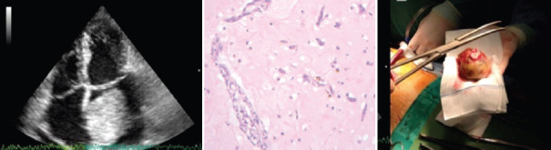
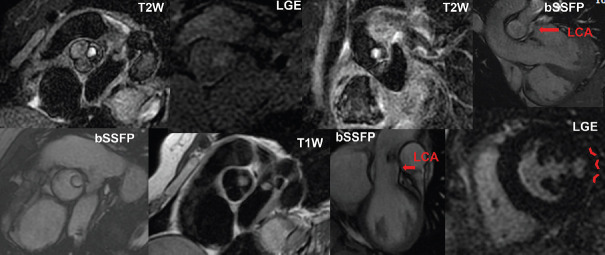
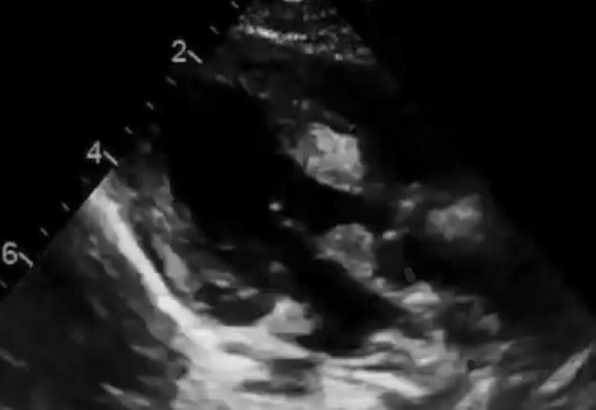
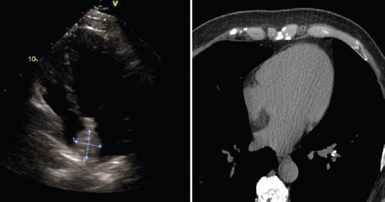
Studying cardiac masses is one of the most challenging tasks for cardiac imagers. The aim of this review article is to focus on the modern imaging of cardiac masses proceeding through the most frequent ones. Cardiac benign masses such as myxoma, cardiac papillary fibroelastoma, rhabdomyoma, lipoma, and hemangioma are browsed considering the usefulness of most common cardiovascular imaging tools, such as ultrasound techniques, cardiac computed tomography, cardiac magnetic resonance, and in the diagnostic process. In the same way, the most frequent malignant cardiac masses, such as angiosarcoma and metastases, are highlighted. Then, the article browses through nontumoral masses such as cysts, mitral caseous degenerative formations, thrombi, and vegetations, highlighting the differential diagnosis between them. In addition, the article helps in recognizing anatomic normal variants that should not be misdiagnosed as pathological entities.

Keywords: Noninvasive cardiovascular imaging; nontumoral cardiac masses; tumoral cardiac masses.


Conflict of interest statement

There are no conflicts of interest.

Figures

Figure 1
Figure 1
Giant LA myxoma. Left panel: TTE 4-C view showing a round shaped and floating LA giant mass (7 cm × 4 cm) with broad attachment to the entire fossa ovalis, protruding into the mitral valve, with an enlarged LA. Central Panel: Histology of the resected myxoma showing amyxomatous tissue with acid-mucopolysaccharide matrix and polygonal cells. Right panel: surgical specimen of the giant resected myxoma (7 Å~ 4 cm) with attached interatrial septum. LA = Left atrium, TTE = Transthoracic echocardiography
Figure 2
Figure 2
MRI imaging of aortic valve papillary fibroelastomas in form of hyperintense rounded structure on the valve. MRI = Magnetic resonance imaging, T1W = T1-weighted imaging, LGE = Late gadolinium enhancement, T2W = T2-weighted imaging, bSSFP = Balanced steady state free precession, LCA = Left coronary artery
Figure 3
Figure 3
Transthoracic echocardiography of left ventricle rhabdomyoma. It appears as multiple homogenous, well circumscribed, hyperechoic intramural masses
Figure 4
Figure 4
Lipomatous hypertrophy of the interatrial septum at transthoracic echocardiography and MRI imaging. MRI = Magnetic resonance imaging
Figure 5
Figure 5
3D trans-esophageal echocardiography showing right atrial angiosarcoma. 3D = Three-dimensional
Figure 6
Figure 6
A pericardial cyst located in the anterior mediastinum at TTE and MRI imaging. TTE = Transthoracic echocardiography, MRI = Magnetic resonance imaging
Figure 7
Figure 7
Giant hydatic cist inside LV at TEE (courtesy of Anita Sadeghpour). LV = Left ventricle, TEE = Transesophageal echocardiography
Figure 8
Figure 8
3D-TEE image of a giant (4 cm × 4 cm) left atrial old and organized thrombus with robust fibrotic coating and central hemolytic area. 3D = Three-dimensional, TEE = Transesophageal echocardiography
Figure 9
Figure 9
Transthoracic and transesophageal echocardiography images showing a right-sided large thrombus through the tricuspid valve
Figure 10
Figure 10
(a) Apical 4-chamber view showing Chiari network within the RA (asterisk). (b) Moderator band (arrow). (c) Pacemaker lead crossing RA and RV (arrows). (d) Parasternal long axis view showing calcification of posterior mitral valve leaflet (asterisk). (e) Reverberation artifact projected into the LV (arrow). LA = Left atrium, LV = Left ventricle, RA = Right atrium, RV = Right ventricle

References

    1. Bianco F, Bucciarelli V, Todiere G, Barison A, Festa P, Ait-Ali L, et al. Cardiac tumors: Role of magnetic resonance imaging. G Ital Cardiol (Rome) 2017;18:286–94. - PubMed
    1. Ha JW, Kang WC, Chung N, Chang BC, Rim SJ, Kwon JW, et al. Echocardiographic and morphologic characteristics of left atrial myxoma and their relation to systemic embolism. Am J Cardiol. 1999;83:1579–82. A8. - PubMed
    1. Yoon DH, Roberts W. Sex distribution in cardiac myxomas. Am J Cardiol. 2002;90:563–5. - PubMed
    1. Wang JG, Wang B, Hu Y, Liu JH, Liu B, Liu H, et al. Clinicopathologic features and outcomes of primary cardiac tumors: A 16-year-experience with 212 patients at a Chinese medical center. Cardiovasc Pathol. 2018;33:45–54. - PubMed
    1. Scalise M, Torella M, Marino F, Ravo M, Giurato G, Vicinanza C, et al. Atrial myxomas arise from multipotent cardiac stem cells. Eur Heart J. 2020;41:4332–45. - PMC - PubMed