

Rapid Objective Testing of Visual Function Matched to the ETDRS Grid and Its Diagnostic Power in Age-Related Macular Degeneration
- PMID: 36249701
- PMCID: PMC9559873
- DOI: 10.1016/j.xops.2022.100143
Rapid Objective Testing of Visual Function Matched to the ETDRS Grid and Its Diagnostic Power in Age-Related Macular Degeneration
Abstract
Purpose: To study the power of an 80-second multifocal pupillographic objective perimetry (mfPOP) test tailored to the ETDRS grid to diagnose age-related macular degeneration (AMD) by Age-Related Eye Disease Study (AREDS) severity grade.
Design: Evaluation of a diagnostic technology.
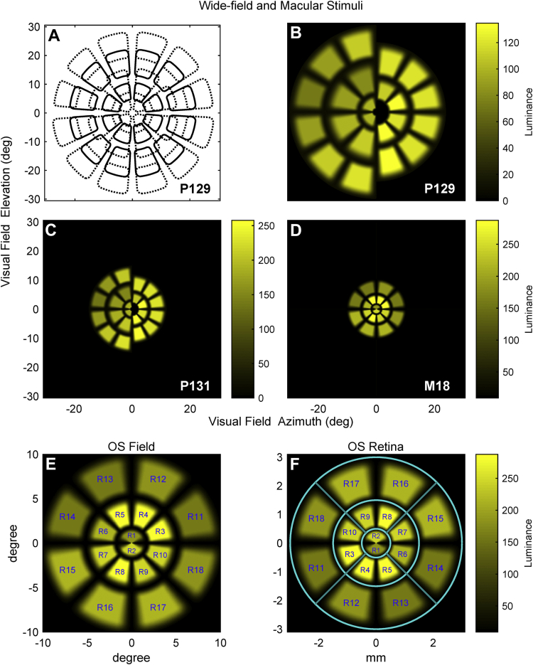
Methods: We compared diagnostic power of acuity, ETDRS grid retinal thickness data, new 80-second M18 mfPOP test, and two wider-field 6-minute mfPOP tests (Macular-P131, Widefield-P129). The M18 stimuli match the size and shape of bifurcated ETDRS grid regions, allowing easy structure-function comparisons. M18, P129, and P131 stimuli test both eyes concurrently. We recruited 34 patients with early-stage AMD with a mean ± standard deviation (SD) age of 72.6 ± 7.06 years. The M18 and P129 plus P131 stimuli had 26 and 51 control participants, respectively with mean ± SD ages of 73.1 ± 8.17 years and 72.1 ± 5.83 years, respectively. Multifocal pupillographic objective perimetry testing used the Food and Drug Administration-cleared Objective FIELD Analyzer (OFA; Konan Medical USA).
Main outcome measures: Percentage area under the receiver operator characteristic curve (AUC) and Hedge's g effect size.
Results: Acuity and OCT ETDRS grid thickness and volume produced reasonable diagnostic power (percentage AUC) for AREDS grade 4 eyes at 83.9 ± 9.98% and 90.2 ± 6.32% (mean ± standard error), respectively, but not for eyes with less severe disease. By contrast, M18 stimuli produced percentage AUCs from 72.8 ± 6.65% (AREDS grade 2) to 92.9 ± 3.93% (AREDS grade 4), and 82.9 ± 3.71% for all eyes. Hedge's g effect sizes ranged from 0.84 to 2.32 (large to huge). Percentage AUC for P131 stimuli performed similarly and for P129 performed somewhat less well.
Conclusions: The rapid and objective M18 test provided diagnostic power comparable with that of wider-field 6-minute mfPOP tests. Unlike acuity or OCT ETDRS grid data, OFA tests produced reasonable diagnostic power in AREDS grade 1 to 3 eyes.
Keywords: AMD, age-related macular degeneration; AREDS, Age-Related Eye Disease Study; AUC, area under the receiver operating characteristic curve; BCVA, best-corrected visual acuity; ETDRS-grid OCT; OFA, Objective FIELD Analyzer; PD, pattern deviation; ROC, receiver operating characteristic; SD, standard deviation; TD, total deviation; abs(DelayDiff), delay difference; macular function; mfPOP, multifocal pupillographic objective perimetry; mfVEP, multifocal visual evoked potential; multifocal pupillography; objective perimetry; rapid perimetry.
© 2022 by the American Academy of Ophthalmology.
Figures

References
-
- Wong W.L., Su X., Li X., et al. Global prevalence of age-related macular degeneration and disease burden projection for 2020 and 2040: a systematic review and meta-analysis. Lancet Glob Health. 2014;2(2):e106–e116. - PubMed
-
- Global Burden of Disease Blindness and Vision Impairment Collaborators Vision Loss Expert Group of the Global Burden of Disease Study. Causes of blindness and vision impairment in 2020 and trends over 30 years, and prevalence of avoidable blindness in relation to VISION 2020: the Right to Sight: an analysis for the Global Burden of Disease Study. Lancet Glob Health. 2021;9(2):e144–e160. - PMC - PubMed
-
- Kawasaki R., Yasuda M., Song S.J., et al. The prevalence of age-related macular degeneration in Asians: a systematic review and meta-analysis. Ophthalmology. 2010;117(5):921–927. - PubMed
LinkOut - more resources
Full Text Sources
