Review
. 2022 Sep 30:9:1015771.
doi: 10.3389/fmolb.2022.1015771. eCollection 2022.

Bacterial glycosylation, it's complicated

Affiliations
Review

Bacterial glycosylation, it's complicated

Christine M Szymanski. Front Mol Biosci. .

Abstract

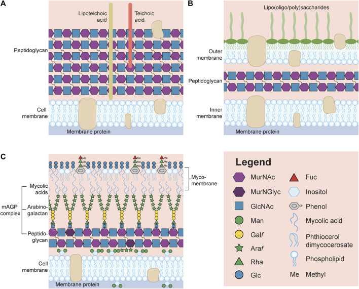
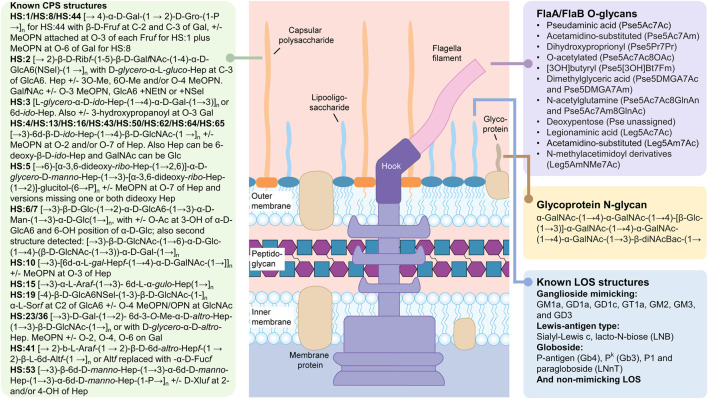
Each microbe has the ability to produce a wide variety of sugar structures that includes some combination of glycolipids, glycoproteins, exopolysaccharides and oligosaccharides. For example, bacteria may synthesize lipooligosaccharides or lipopolysaccharides, teichoic and lipoteichoic acids, N- and O-linked glycoproteins, capsular polysaccharides, exopolysaccharides, poly-N-acetylglycosamine polymers, peptidoglycans, osmoregulated periplasmic glucans, trehalose or glycogen, just to name a few of the more broadly distributed carbohydrates that have been studied. The composition of many of these glycans are typically dissimilar from those described in eukaryotes, both in the seemingly endless repertoire of sugars that microbes are capable of synthesizing, and in the unique modifications that are attached to the carbohydrate residues. Furthermore, strain-to-strain differences in the carbohydrate building blocks used to create these glycoconjugates are the norm, and many strains possess additional mechanisms for turning on and off transferases that add specific monosaccharides and/or modifications, exponentially contributing to the structural heterogeneity observed by a single isolate, and preventing any structural generalization at the species level. In the past, a greater proportion of research effort was directed toward characterizing human pathogens rather than commensals or environmental isolates, and historically, the focus was on microbes that were simple to grow in large quantities and straightforward to genetically manipulate. These studies have revealed the complexity that exists among individual strains and have formed a foundation to better understand how other microbes, hosts and environments further transform the glycan composition of a single isolate. These studies also motivate researchers to further explore microbial glycan diversity, particularly as more sensitive analytical instruments and methods are developed to examine microbial populations in situ rather than in large scale from an enriched nutrient flask. This review emphasizes many of these points using the common foodborne pathogen Campylobacter jejuni as the model microbe.

Keywords: Campylobacter jejuni; carbohydrates; glycoconjugates; phase-variation; polysaccharides.


Conflict of interest statement

The author declares that the research was conducted in the absence of any commercial or financial relationships that could be construed as a potential conflict of interest.

Figures

FIGURE 1
FIGURE 1
General representation of the surface of a Gram-positive (A) and Gram-negative (B) bacterium. (C) Schematic of the surface of Mycobacterium tuberculosis unable to be identified by the Gram stain. Note that many structures, including capsular polysaccharides and glycoproteins, have been omitted in order to reduce the complexity of the image. Also, lipoteichoic acids, teichoic acids and lipooligosaccharides/lipopolysaccharides are not drawn with the symbol nomenclature for glycans (SNFG, https://www.ncbi.nlm.nih.gov/glycans/snfg.html) due to the limitless structures that could be drawn for each glycoconjugate. MurNAc, N-acetylmuramic acid; MurNGlyc, N-glycolylmuramic acid; GlcNAc, N-acetylglucosamine; Man, mannose; Galf, galactofuranose; Araf, arabinofuranose; Rha, rhamnose; Glc, glucose; Fuc, fucose.
FIGURE 2
FIGURE 2
Carbohydrate structures synthesized by Campylobacter jejuni. The summarized structures include the capsular polysaccharides (CPS), the lipooligosaccharides (LOS) and the N- and O-linked glycoprotein glycans. N-glycans modify a large number of glycoproteins while O-glycans are found exclusively on the FlaA and FlaB subunits of the flagellum. Peptidoglycan sugars are the same as described in Figure 1. The free oligosaccharides (fOS) derived from the N-glycan pathway are not shown (see text). All sugars are in the pyranose form unless otherwise noted. Pse5Ac7Ac, pseudaminic acid (5,7-diacetamido-3,5,7,9-tetradeoxy-L-glycero-α-L-manno nonulosonic acid; Leg5Ac7Ac), legionaminic acid (5,7-diacetamido-3,5,7,9-tetradeoxy-D-glycero-β-D-galacto-nonulosonic acid); Fm, formyl; Gal, galactose; Gro, glycerol; Fruf, fructose in furanose configuration; MeOPN, methyl phosphoramidate; Rib, ribose; GalNAc, N-acetylgalactosamine; GlcA6, glucuronic acid; Hep, heptose; GlcNAc, N-acetylglucosamine; Glc, glucose; Man, mannose; Ara, arabinose; Alt, altrose; Sel, serinol; Xlu, xylulose. Structures were found in: (McNally et al., 2005; McNally et al., 2006; Hitchen et al., 2010; Houliston et al., 2011; Monteiro et al., 2018; Riegert et al., 2021).
FIGURE 3
FIGURE 3
(A) Schematic demonstrating the reported capsular polysaccharide (CPS) structures originating from Campylobacter jejuni strain NCTC 11168, from ~ 1000 possible structures, using the Symbol Nomenclature for Glycans with some modifications. The CPS structure shown is the [→ 2)-β-D-Ribf-(1–5)-β-D-GalfNAc-(1–4)-α-D-GlcA6(NSel)-(1 →]n repeating unit with D-glycero-α-L-gluco-Hep (blue hexagon) at C-3 of GlcA (diamond). The Hep can be further modified+/-3O-Me, 6O-Me (methyl groups are grey circles) and/or O-4 MeOPN (methyl phosphoramidate drawn as red circle). In addition, the GalfNAc (yellow square with f to designate furanose form) can be modified at O-3+/-MeOPN, and GlcA6 can be modified with either NSel (N-serinol, green oval) or NEtN (N-ethanolamine, purple rectangle). Ribose is shown as the pink star. (B) Original image from Szymanski et al., 2003 showing differences in CPS structures (c-f), and their corresponding silver-staining (a) and immunoreactivity (b) from single colony isolates originating from same culture. (a) Silver-stained deoxycholate-PAGE and (b) western blot detected with HS:2 typing sera both showing: lane 1, NCTC 11168 wildtype population; lane 2, 11168 variant one; lane 3, 11168 variant two; and lane 4, 11168 variant 3. (c-f) High resolution magic angle spinning (HR-MAS) NMR spectra of the wildtype population (c), variant 1 with arrow indicating ethanolamine resonance (d), variant 2 with arrow indicating MeOPN modification identified for the first time (e), and variant 3 with the arrow indicating loss of OMe resonance (f). Anomeric resonances in c-f are labeled A (Ribf), B (GlcA6), C (GalfNAc), and D (Hep). See (Szymanski et al., 2003) for more information.

References

    1. Abrahams K. A., Besra G. S. (2018). Mycobacterial cell wall biosynthesis: a multifaceted antibiotic target. Parasitology 145 (2), 116–133. 10.1017/S0031182016002377 - DOI - PMC - PubMed
    1. Amour C., Gratz J., Mduma E., Svensen E., Rogawski E. T., McGrath M., et al. (2016). Epidemiology and impact of Campylobacter infection in children in 8 low-resource settings: results from the MAL-ED study. Clin. Infect. Dis. 63 (9), 1171–1179. 10.1093/cid/ciw542 - DOI - PMC - PubMed
    1. Batt S. M., Minnikin D. E., Besra G. S. (2020). The thick waxy coat of mycobacteria, a protective layer against antibiotics and the host's immune system. Biochem. J. 477 (10), 1983–2006. 10.1042/BCJ20200194 - DOI - PMC - PubMed
    1. Bian X., Garber J. M., Cooper K. K., Huynh S., Jones J., Mills M. K., et al. (2020). Campylobacter abundance in breastfed infants and identification of a new species in the global enterics multicenter study. mSphere 5 (1), 007355–e819. 10.1128/mSphere.00735-19 - DOI - PMC - PubMed
    1. Brockhausen I. (2014). Crossroads between bacterial and mammalian glycosyltransferases. Front. Immunol. 5, 492. 10.3389/fimmu.2014.00492 - DOI - PMC - PubMed

LinkOut - more resources