. 2022 Dec 13:102:adv00834.
doi: 10.2340/actadv.v102.325.

Longitudinal Evaluation of Biomarkers in Wound Fluids from Venous Leg Ulcers and Split-thickness Skin Graft Donor Site Wounds Treated with a Protease-modulating Wound Dressing

Affiliations

Longitudinal Evaluation of Biomarkers in Wound Fluids from Venous Leg Ulcers and Split-thickness Skin Graft Donor Site Wounds Treated with a Protease-modulating Wound Dressing

Jacek Mikosiński et al. Acta Derm Venereol. .

Abstract

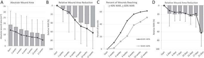
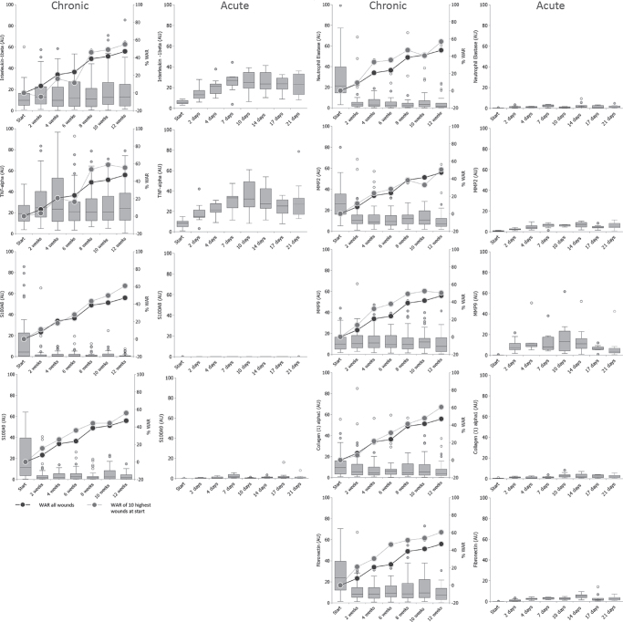
Venous leg ulcers represent a clinical challenge and impair the quality of life of patients. This study examines impaired wound healing in venous leg ulcers at the molecular level. Protein expression patterns for biomarkers were analysed in venous leg ulcer wound fluids from 57 patients treated with a protease-modulating polyacrylate wound dressing for 12 weeks, and compared with exudates from 10 acute split-thickness wounds. Wound healing improved in the venous leg ulcer wounds: 61.4% of the 57 patients with venous leg ulcer achieved a relative wound area reduction of ≥ 40%, and 50.9% of the total 57 patients achieved a relative wound area reduction of ≥ 60%. Within the first 14 days, abundances of S100A8, S100A9, neutrophil elastase, matrix metalloproteinase-2, and fibronectin in venous leg ulcer exudates decreased significantly and remained stable, yet higher than in acute wounds. Interleukin-1β, tumour necrosis factor alpha, and matrix metalloproteinase-9 abundance ranges were similar in venous leg ulcers and acute wound fluids. Collagen (I) α1 abundance was higher in venous leg ulcer wound fluids and was not significantly regulated. Overall, significant biomarker changes occurred in the first 14 days before a clinically robust healing response in the venous leg ulcer cohort.


Conflict of interest statement

Conflicts of interest: HS is a full-time employee of Hartmann. The other authors have no conflicts of interest to declare.

Figures

Fig. 1
Fig. 1
Wound area development in the venous leg ulcer cohort. (A) Development of the absolute wound area (in cm2). (B) Relative wound area reduction (WAR) (in %). (C) Percentage of wounds achieving a relative WAR of ≥40% (dark dots) and ≥60% (grey dots). (D) Relative wound area reduction (WAR) (in %) in the split-thickness donor site wounds. (A–D) Mean values are shown in grey boxes with the corresponding median values as dark dots.
Fig. 2
Fig. 2
Evolution of inflammatory cytokine concentrations in venous leg ulcers (VLUs) (chronic) and split-thickness wounds (acute). Grey boxes indicate cytokine concentrations in arbitrary units (AU). Superimposed dark grey dots indicate the relative wound area reduction (WAR) development of the VLU wounds for the whole population. Light grey dots show the relative WAR was followed over time in 10 wounds with the highest respective biomarker abundance at the start.
Fig. 3
Fig. 3
Biomarker profiles of the venous leg ulcer wound fluids. Grey areas represent the mean values of the whole population; dark lines indicate the means of the biomarker of the 10 patients with the poorest performance or the relative wound area reduction (WAR) at the respective time-points. (A) At the start, (B) 2 weeks, (C) 4 weeks, (D) 6 weeks, (E) 8 weeks, (F) 10 weeks and (G) 12 weeks. As for the presentation of the 10 poorest responders, the calculation of the relative WAR development was not possible; the relative WAR values of the 2-week time-point were used to rank the biomarker responses at the start. (H) Expression profiles of the acute wound healing cohort at 7 days. IL-1b; interleukin 1b; TNF-a: tumour necrosis factor-alpha, ELNE: neutrophil elastase; MMP2: matrix metalloprotease-2; MMP9: matrix metalloprotease-9; Coll (I) a1: collagen (I) a1; FN: fibronectin.

References

    1. Patton D, Avsar P, Sayeh A, Budri A, O’Connor T, Walsh S, et al. . A meta-review of the impact of compression therapy on venous leg ulcer healing. Int Wound J 2022; Jul 18. [Online ahead of print]. - PMC - PubMed
    1. O’Meara S, Cullum N, Nelson EA, Dumville JC. Compression for venous leg ulcers. Cochrane Database Syst Rev 2012; 11: CD000265. - PMC - PubMed
    1. Marston WA, Ennis WJ, Lantis JC, Kirsner RS, Galiano RD, Vanscheidt W, et al. . Baseline factors affecting closure of venous leg ulcers. J Vasc Surg Venous Lymphat Disord 2017; 5: 829–835.e1. - PubMed
    1. Kantor J, Margolis DJ. A multicentre study of percentage change in venous leg ulcer area as a prognostic index of healing at 24 weeks. Br J Dermatol 2000; 142: 960–964. - PubMed
    1. Guest JF, Fuller GW, Vowden P. Venous leg ulcer management in clinical practice in the UK: costs and outcomes. Int Wound J 2018; 15: 29–37. - PMC - PubMed

Substances