

Intraoperative determination of the risky angles and safe distances for preventing deep femoral artery injury during proximal femoral nailing for hip fractures in Asian people
- PMID: 36250880
- PMCID: PMC9682588
- DOI: 10.5152/j.aott.2022.22061
Intraoperative determination of the risky angles and safe distances for preventing deep femoral artery injury during proximal femoral nailing for hip fractures in Asian people
Abstract
Objective: During proximal femoral nailing, deep femoral artery injury, a rare condition, is often missed and found late, leading to intractable complications such as false aneurysm, hematoma, and anemia. We aimed to determine the novel indicators of the high-risk vertical range and axial angle for deep femoral artery injury that can be easily confirmed intraoperatively using fluoroscopy for hip fracture.
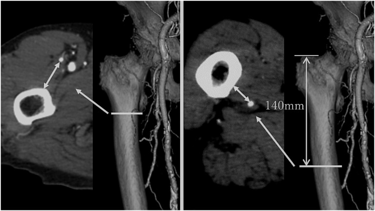
Methods: In a single hospital, the lower extremity computed tomography angiographies of 88 patients (50 men and 38 women) were analyzed. A reference plane was defined as the femoral neck and shaft on the same straight line in the lateral view. Reference points were the lower end of the lesser trochanter and distal femur at 140 mm from the tip of the greater trochanter. To determine the high-risk angle for deep femoral artery injury based on the reference plane, the angle from the reference plane to the deep femoral artery (bone-arterial angle) and the shortest distance between the surfaces of the femur and the deep femoral artery (bone-artery distance) were measured at the lesser trochanter and the greater trochanter. We analyzed the bone-arterial angle and bone-artery distance values, their differences among the sexes, and their correlation with body height and body weight.
Results: Overall, in the lesser trochanter, the mean bone-arterial angle and bone-artery distance were 19.2° ± 8.0° and 22.9 ± 4.7 mm, respectively. In the greater trochanter, the mean bone-arterial angle and bone-artery distance were -33.9° ± 17.0° and 11.3 ± 4.1 mm, respectively. The mean bone-artery distance of the lesser trochanter was significantly longer in men than in women (24.1 ± 4.5 mm and 21.4 ± 4.5 mm, respectively, P < 0.01), and for the lesser trochanter, positive correlations were found between body height and both bone- arterial angle and bone-artery distance (r=0.373, P < 0.001; and r=0.456, P < 0.0001, respectively), with body weight and bone-artery distance positively correlated (r=0.367, P < 0.001). At the greater trochanter, there were negative correlations between body height and bone-arterial angle (r=-0.5671, P < 0.0001), body weight, and bone-arterial angle (r=-0.338, P < 0.01).
Conclusion: The knowledge of our reference plane and high-risk angles and distances allows surgeons to minimize the risk of deep femoral artery injury. These are easily confirmed intraoperatively using fluoroscopy, allowing surgeons to avoid maneuvering in the deep femoral artery range.
Level of evidence: Level IV, Diagnostic Study.
Figures


Similar articles
-
Evaluation of the positional relationship between femoral arteries and distal screws in the proximal femoral intramedullary nail for preventing iatrogenic vascular injury.Injury. 2020 Feb;51(2):384-388. doi: 10.1016/j.injury.2019.10.003. Epub 2019 Oct 3. Injury. 2020. PMID: 31668355
-
Risk of Arterial Injury During Hip Internal Fixation.J Bone Joint Surg Am. 2019 Nov 6;101(21):1961-1964. doi: 10.2106/JBJS.19.00256. J Bone Joint Surg Am. 2019. PMID: 31596820
-
Is the Lesser Trochanter Profile a Reliable Means of Restoring Anatomic Rotation After Femur Fracture Fixation?Clin Orthop Relat Res. 2018 Jun;476(6):1253-1261. doi: 10.1007/s11999.0000000000000226. Clin Orthop Relat Res. 2018. PMID: 29470236 Free PMC article.
-
Delayed femoral artery injury caused by heterotopic ossification: a rare case report and review of the literature.BMC Musculoskelet Disord. 2024 Jun 20;25(1):485. doi: 10.1186/s12891-024-07609-5. BMC Musculoskelet Disord. 2024. PMID: 38902664 Free PMC article. Review.
-
Secondary profunda femoris artery injury after intramedullary femoral nailing in a geriatric pertrochanteric femur fracture: case report.Eur J Orthop Surg Traumatol. 2019 Dec;29(8):1811-1814. doi: 10.1007/s00590-019-02500-9. Epub 2019 Jul 26. Eur J Orthop Surg Traumatol. 2019. PMID: 31346724 Review.
Cited by
-
Iatrogenic injury to the descending branch of the lateral circumflex femoral artery during intertrochanteric fracture fixation: a case report of guide pin-related vascular complication.Acta Orthop Traumatol Turc. 2025 Jul 18;59(4):237-240. doi: 10.5152/j.aott.2025.24071. Acta Orthop Traumatol Turc. 2025. PMID: 40728093 Free PMC article.
References
-
- Potenza V, Saputo U, Catellani F, Farsetti P, Caterini R. Laceration of a branch of the profunda femoris artery caused by a spike of the displaced lesser trochanter in an inter-trochanteric femoral fracture. A case report. Int J Surg Case Rep. 2016;24:195 198. 10.1016/j.ijscr.2016.05.048) - DOI - PMC - PubMed
MeSH terms
LinkOut - more resources
Full Text Sources
Medical
Research Materials
