. 2022 Oct 17;18(10):e1010300.
doi: 10.1371/journal.pgen.1010300. eCollection 2022 Oct.

RaScALL: Rapid (Ra) screening (Sc) of RNA-seq data for prognostically significant genomic alterations in acute lymphoblastic leukaemia (ALL)

Affiliations

RaScALL: Rapid (Ra) screening (Sc) of RNA-seq data for prognostically significant genomic alterations in acute lymphoblastic leukaemia (ALL)

Jacqueline Rehn et al. PLoS Genet. .

Abstract

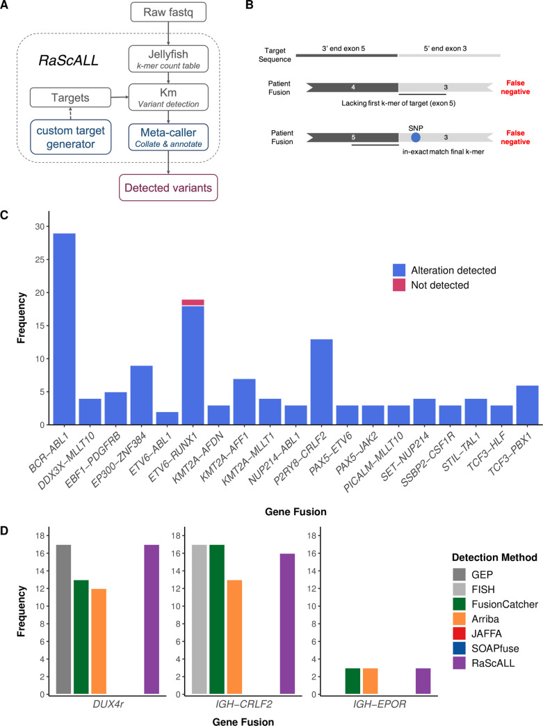
RNA-sequencing (RNA-seq) efforts in acute lymphoblastic leukaemia (ALL) have identified numerous prognostically significant genomic alterations which can guide diagnostic risk stratification and treatment choices when detected early. However, integrating RNA-seq in a clinical setting requires rapid detection and accurate reporting of clinically relevant alterations. Here we present RaScALL, an implementation of the k-mer based variant detection tool km, capable of identifying more than 100 prognostically significant lesions observed in ALL, including gene fusions, single nucleotide variants and focal gene deletions. We compared genomic alterations detected by RaScALL and those reported by alignment-based de novo variant detection tools in a study cohort of 180 Australian patient samples. Results were validated using 100 patient samples from a published North American cohort. RaScALL demonstrated a high degree of accuracy for reporting subtype defining genomic alterations. Gene fusions, including difficult to detect fusions involving EPOR and DUX4, were accurately identified in 98% of reported cases in the study cohort (n = 164) and 95% of samples (n = 63) in the validation cohort. Pathogenic sequence variants were correctly identified in 75% of tested samples, including all cases involving subtype defining variants PAX5 p.P80R (n = 12) and IKZF1 p.N159Y (n = 4). Intragenic IKZF1 deletions resulting in aberrant transcript isoforms were also detectable with 98% accuracy. Importantly, the median analysis time for detection of all targeted alterations averaged 22 minutes per sample, significantly shorter than standard alignment-based approaches. The application of RaScALL enables rapid identification and reporting of previously identified genomic alterations of known clinical relevance.


Conflict of interest statement

The authors have declared that no competing interests exist.

Figures

Fig 1
Fig 1. Genomic alterations for patients within the study cohort.
Reported genomic alterations for 180 ALL samples analysed by RNA-seq, including 160 B-ALL patient samples (left panel), 18 patients samples classified as T-ALL (right panel), and two samples with acute undifferentiated leukaemia or acute bi-phenotypic leukaemia (middle panel). Each column represents a single patient. Patients were classified into a recognised subtype based on the detected driver mutation. For each patient, the deletion status of IKZF1, as reported by multiplex ligation-dependent probe amplification, as well as patient diagnosis, age group and sample timepoint are shown and coloured according to the key. Clinically relevant gene fusions detected with RNA-seq are indicated in the middle panel and shown in dark grey. The bottom panel indicates samples for which known pathogenic SNVs were detected by GATK Haplotype caller (shown in dark grey).
Fig 2
Fig 2. Detection of subtype-defining gene fusions by RaScALL.
(A) RaScALL workflow. RaScALL is a self-contained command line utility for detecting ALL specific variants. RNA-seq data in the form of raw FASTQ files are indexed by jellyfish to generate a k-mer count table. Next, gene fusions, focal gene deletions, SNVs and InDels are detected with km for genomic regions specified in the target set and the output is filtered to provide a single file with clinically relevant alterations for reporting. (B) Schematic representation of gene fusion target sequences (top panel) and causes of false-negative reports. A false negative occur when the patient possesses an alternate breakpoint lacking one of the exons in the gene fusion target (middle panel), or due to the presence of a SNP preventing an exact match of the first or final k-mer in the target sequence (bottom panel). (C) Frequency of detection of non-IGH gene fusions by RaScALL in the study cohort. Blue bar indicating that RaScALL detected the same gene fusion reported by two or more alignment-based fusion calling algorithms and a single sample in pink for which RaScALL failed to detect a clinically reported gene fusion. (D) Frequency of IGH-CRLF2, IGH-EPOR or DUX4r in the study cohort as determined by FISH for IGH-CRLF2 or gene expression profiling (GEP) for DUX4r (grey). Coloured bars indicate the number of samples for which RaScALL (purple) or one of 4 alignment-based fusion calling algorithms detected the rearrangement in the given patient sample.
Fig 3
Fig 3. Detection of IKZF1 intragenic deletions with RaScALL.
Samples were analysed by RaScALL for the presence of target sequences representing IKZF1 Δ2–3, Δ2–7 and Δ4–7 deletions. Deletion events reported by RaScALL were then compared to IKZF1 deletions reported by MLPA analysis using P335 and P202 kits. (A) Minimum coverage (k-mers) across target sequences (log10 scale) for targets representing IKZF1 Δ2–3 deletion, Δ2–7 deletion or Δ4–7 deletion. Points are coloured concordant (blue) when the same IKZF1 deletion was identified by MLPA or discordant (pink) when MLPA reported no IKZF1 deletion for the sample. (B) Sample frequency for IKZF1 status reported as either no deletion, Δ2–3 deletion, Δ2–7 deletion or Δ4–7 IKZF1 deletion (minimum k-mer coverage > = 10). Bars are coloured according to whether the IKZF1 deletion reported by RaScALL was supported (Concordant, dark blue), or not supported (discordant, pink) by MLPA analysis.
Fig 4
Fig 4. RaScALL accurately detects subtype defining genetic variants in a validation cohort of 100 B-ALL patient samples.
Fusions (A) and variants (B) detected by RaScALL and their concordance with clinically reported alterations. The vertical axis indicates the number of detected alterations while the horizontal axis indicates the specific gene fusion (A) or gene in which the variant occurred (B). Concordant results are indicated in blue and discordant indicated in dark red for false negative results and light red for false positive results. (C) Runtimes for RaScALL in minutes. Wall clock time for 100 samples represented as boxplots. The vertical axis indicates the separate run times for indexing of raw data and assessment of variants with different target sets. The Number of targets in each set is indicated.

References

    1. Coccaro N, Anelli L, Zagaria A, Specchia G, Albano F. Next-Generation Sequencing in Acute Lymphoblastic Leukemia. Int J Mol Sci [Internet]. 2019. Jun 15;20(12). Available from: doi: 10.3390/ijms20122929 - DOI - PMC - PubMed
    1. Malard F, Mohty M. Acute lymphoblastic leukaemia. Lancet. 2020. Apr 4;395(10230):1146–62. doi: 10.1016/S0140-6736(19)33018-1 - DOI - PubMed
    1. Yeung DTO, Osborn MP, White DL. B-cell acute lymphoblastic leukaemia: recent discoveries in molecular pathology, their prognostic significance, and a review of the current classification. Br J Haematol. 2022. Apr;197(1):13–27. doi: 10.1111/bjh.17879 - DOI - PubMed
    1. Li JF, Dai YT, Lilljebjörn H, Shen SH, Cui BW, Bai L, et al.. Transcriptional landscape of B cell precursor acute lymphoblastic leukemia based on an international study of 1,223 cases. Proc Natl Acad Sci U S A [Internet]. 2018; Available from: 10.1073/pnas.1814397115 - DOI - PMC - PubMed
    1. Gu Z, Churchman ML, Roberts KG, Moore I, Zhou X, Nakitandwe J, et al.. PAX5-driven subtypes of B-progenitor acute lymphoblastic leukemia. Nat Genet. 2019. Feb 14;51(2):296–307. doi: 10.1038/s41588-018-0315-5 - DOI - PMC - PubMed

Publication types