. 2022 Nov 1;94(43):14827-14834.
doi: 10.1021/acs.analchem.2c00878. Epub 2022 Oct 17.

LANCE: a Label-Free Live Apoptotic and Necrotic Cell Explorer Using Convolutional Neural Network Image Analysis

Affiliations

LANCE: a Label-Free Live Apoptotic and Necrotic Cell Explorer Using Convolutional Neural Network Image Analysis

Emma B Hartnett et al. Anal Chem. .

Abstract

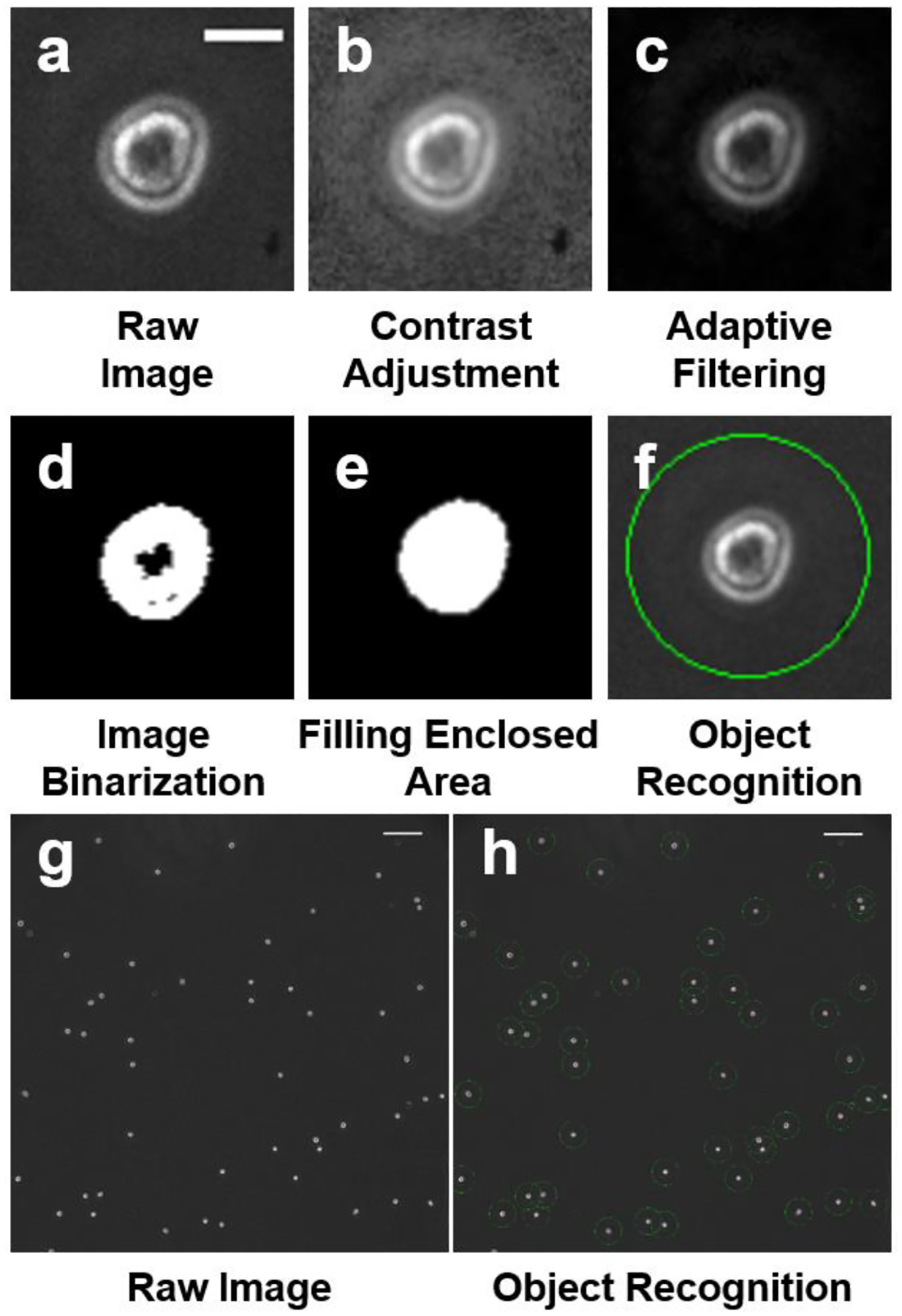
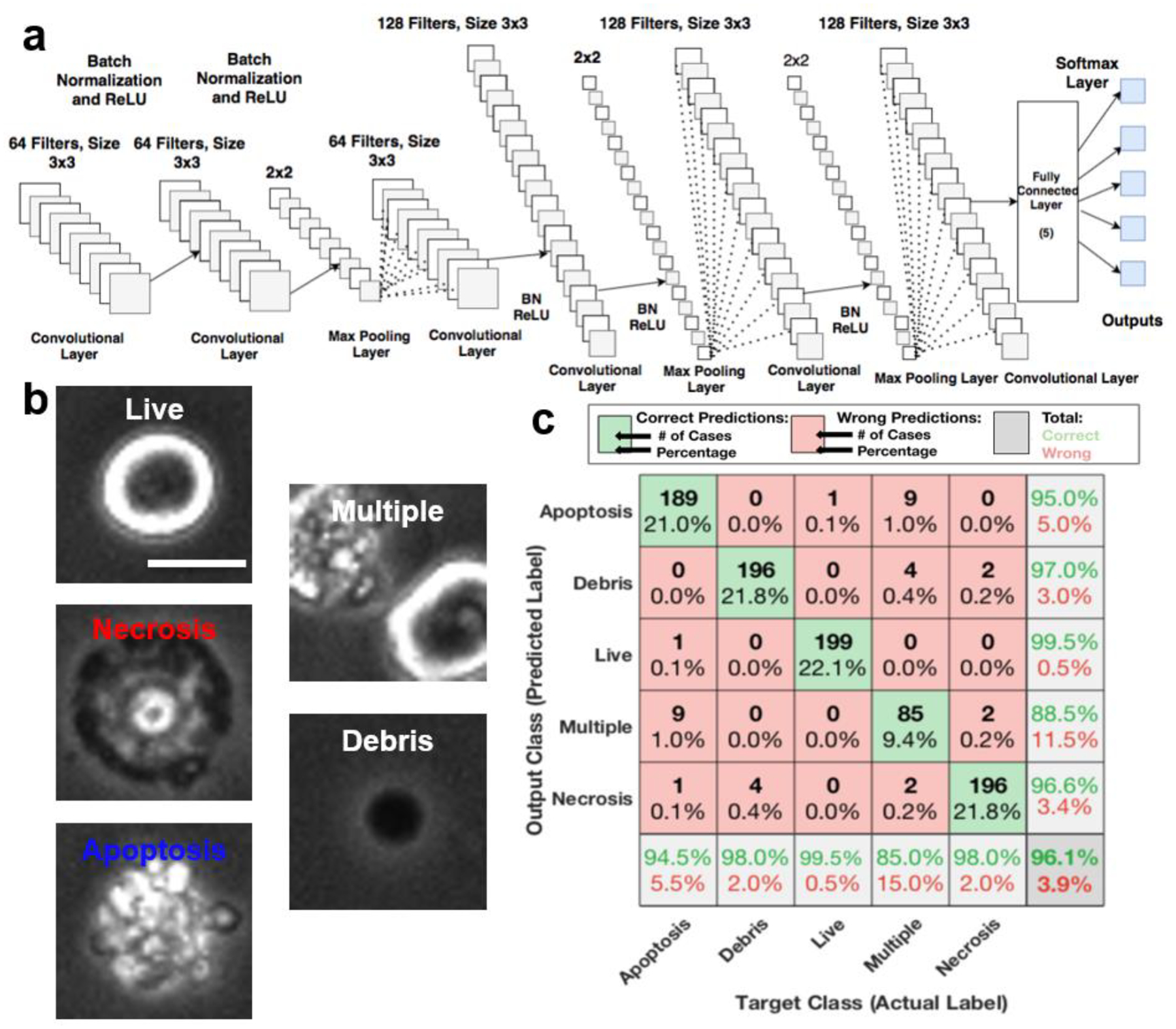
Identifying and quantifying cell death is the basis for all cell death research. Current methods for obtaining these quantitative measurements rely on established biomarkers, yet the marker-based approach suffers from limited marker specificity, high cost of reagents, lengthy sample preparation, and fluorescence imaging. Based on the morphological difference, we developed a Live, Apoptotic, and Necrotic Cell Explorer (LANCE) to categorize cell death status in a label-free manner, by incorporating machine learning and image processing. The LANCE workflow includes cropping individual cells from microscopic images having hundreds of cells, formation of an image database of around 5000 events, training and validation of the convolutional neural network models using multiple cell lines, and treatment conditions. With LANCE, we precisely categorized live, apoptotic, and necrotic cells with a high accuracy of 96.3 ± 0.5%. More importantly, the nondestructive label-free LANCE method allows for tracking time dynamics of the cell death process, which enhances the understanding of subtle cell death regulation at the molecular level. Hence, LANCE is a fast, low-cost, and nondestructive label-free method to distinguish cell status, which can be applied to cell death studies as well as many other biomedical applications.


Conflict of interest statement

COMPETING FINANCIAL INTERESTS

The authors declare no competing financial interests.

Figures

Fig. 1.
Fig. 1.. Cell cropping process from a microscopy image.
(a-f) Image processing and cropping of a single cell. (a) Unaltered raw image. (Scale bar: 13 μm). (b) Contrast adjustment for improving binarization. (c) Adaptive filtering through a 5 pixel by 5-pixel window. (d) Image binarization by a threshold. (e) Filling of an enclosed area. Areas smaller than 150 pixels were eliminated. (f) Cell cropping based on the calculated centroid. Processed images were cropped into 40×40 pixel images, (g) A raw microscopy image (scale bar: 130 μm). (h) A processed image showing all objects were successfully recognized as marked. (Scale bar: 130 μm).
Fig. 2.
Fig. 2.. LANCE prediction of cell status by a convolutional neural network (CNN).
(a) CNN structure characterized by six convolutional layers. (b) Representative images of cell categories (‘Live,’ ‘Apoptosis,’ ‘Necrosis,’ ‘Debris,’ and ‘Multiple’) used in training and prediction (scale bar: 10 μm). (c) Confusion matrix of predicting testing dataset using LANCE demonstrates a high accuracy of 96.1%.
Fig. 3.
Fig. 3.. Comparison between four different network structures.
Error bar represents the standard deviation of 5 independent trials.
Fig. 4.
Fig. 4.. Comparison between images taken by different microscopes.
The length of bars represents the percentage of cell subsets. Error bar represents the standard deviation (N = 4 for Nikon Ti2E, and N = 5 for BioTek Lionheart FX).
Fig. 5.
Fig. 5.. Prediction of the status of iMACs and Jurkat cells.
TNF+CHX was used to induce apoptosis. LPS+OXO or TCZ was used to induce necrosis. The length of bars represents the percentage of cell subsets. Error bar represents the standard deviation of 4–20 images.
Fig. 6.
Fig. 6.. Western blot of GSDME expression and cleavage in U937 and U937-GSDME cells
upon 150 KJ/cm2 UV irradiation (UV) with or without TNF (T) for ~2 hr.
Fig. 7.
Fig. 7.. Time dynamics of cell death.
(a-b) Temporal tracking of U937 (GSDME silenced) and U937-GE (GSDME expressed) cells irradiated by 150 KJ/cm2 UV. Cells were imaged every 30 minutes for 4 hours. Error bar represents the standard deviation of 4 images at each time point. (c) Representative initial image of U937 cells. (d) Representative images of U937 cells four hours after UV irradiation. (e) Representative initial image of U937-GE cells. (f) Representative images of U937-GE cells four hours after UV irradiation. Scale bar: 50 μm.

References

    1. Green DR The Coming Decade of Cell Death Research: Five Riddles. Cell. 2019, 177, 1094–1107, DOI: 10.1016/j.cell.2019.04.024. - DOI - PMC - PubMed
    1. Green DR A BH3 Mimetic for Killing Cancer Cells. Cell. 2016, 165, 1560, DOI: 10.1016/j.cell.2016.05.080. - DOI - PubMed
    1. Galluzzi L; et al. Molecular mechanisms of cell death: recommendations of the Nomenclature Committee on Cell Death 2018. Cell Death Differ. 2018, 25, 486–54, DOI: 10.1038/s41418-017-0012-4. - DOI - PMC - PubMed
    1. Martin SJ; Reutelingsperger CP; McGahon AJ; Rader JA; van Schie RC; LaFace DM; Green DR Early redistribution of plasma membrane phosphatidylserine is a general feature of apoptosis regardless of the initiating stimulus: inhibition by overexpression of Bcl-2 and Abl. J. Exp. Med 1995, 182, 1545–1556, DOI: 10.1084/jem.182.5.1545. - DOI - PMC - PubMed
    1. Gong YN; Guy C; Olauson H; Becker JU; Yang M; Fitzgerald P; Linkermann A; Green DR ESCRT-III Acts Downstream of MLKL to Regulate Necroptotic Cell Death and Its Consequences. Cell. 2017, 169, 286–300 216, DOI: 10.1016/j.cell.2017.03.020. - DOI - PMC - PubMed

Publication types