. 2022 Oct 25;119(43):e2200215119.
doi: 10.1073/pnas.2200215119. Epub 2022 Oct 17.

UBR2 targets myosin heavy chain IIb and IIx for degradation: Molecular mechanism essential for cancer-induced muscle wasting

Affiliations

UBR2 targets myosin heavy chain IIb and IIx for degradation: Molecular mechanism essential for cancer-induced muscle wasting

Song Gao et al. Proc Natl Acad Sci U S A. .

Abstract

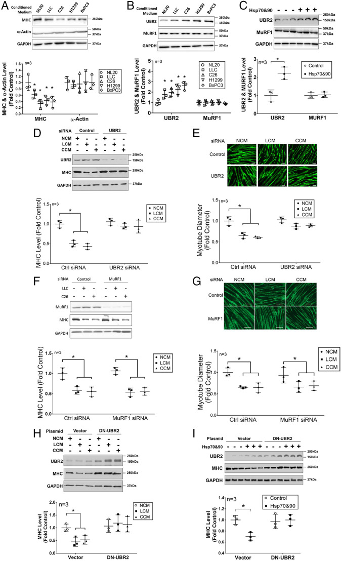
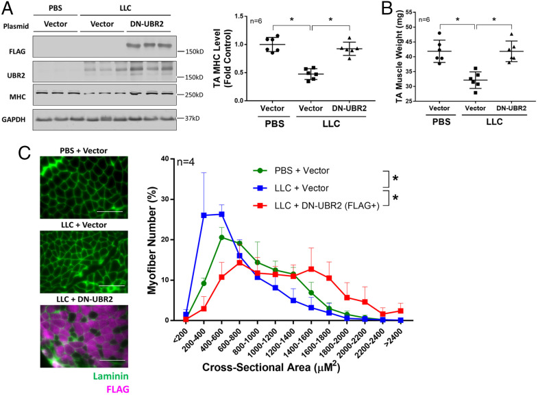
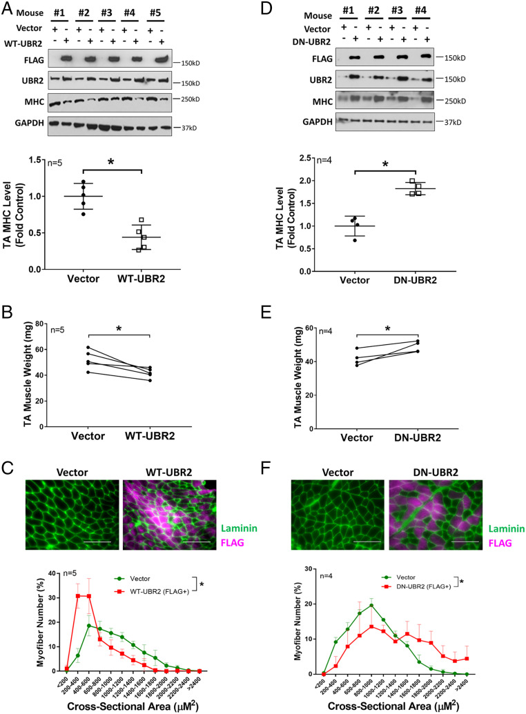
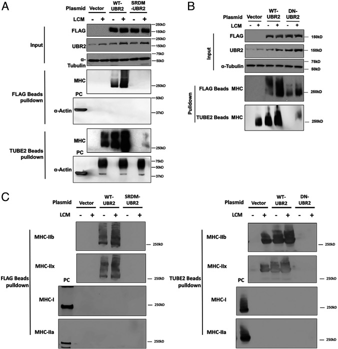
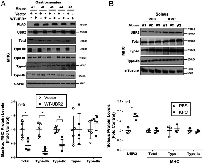
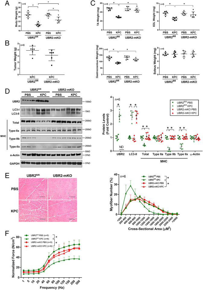
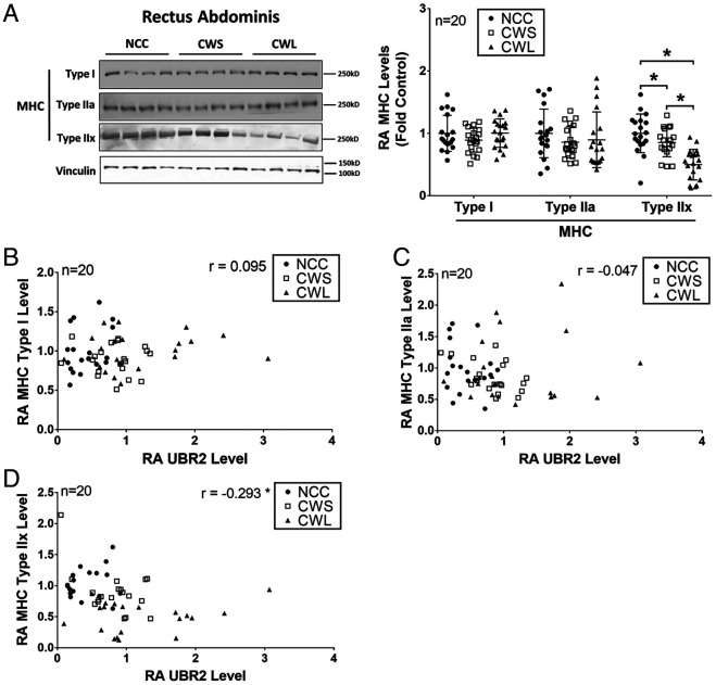
Cancer cachexia is a lethal metabolic syndrome featuring muscle wasting with preferential loss of fast-twitching muscle mass through an undefined mechanism. Here, we show that cancer induces muscle wasting by selectively degrading myosin heavy chain (MHC) subtypes IIb and IIx through E3 ligase UBR2-mediated ubiquitylation. Induction of MHC loss and atrophy in C2C12 myotubes and mouse tibialis anterior (TA) by murine cancer cells required UBR2 up-regulation by cancer. Genetic gain or loss of UBR2 function inversely altered MHC level and muscle mass in TA of tumor-free mice. UBR2 selectively interacted with and ubiquitylated MHC-IIb and MHC-IIx through its substrate recognition and catalytic domain, respectively, in C2C12 myotubes. Elevation of UBR2 in muscle of tumor-bearing or free mice caused loss of MHC-IIb and MHC-IIx but not MHC-I and MHC-IIa or other myofibrillar proteins, including α-actin, troponin, tropomyosin, and tropomodulin. Muscle-specific knockout of UBR2 spared KPC tumor-bearing mice from losing MHC-IIb and MHC-IIx, fast-twitching muscle mass, cross-sectional area, and contractile force. The rectus abdominis (RA) muscle of patients with cachexia-prone cancers displayed a selective reduction of MHC-IIx in correlation with higher UBR2 levels. These data suggest that UBR2 is a regulator of MHC-IIb/IIx essential for cancer-induced muscle wasting, and that therapeutic interventions can be designed by blocking UBR2 up-regulation by cancer.

Keywords: MHC-IIb; MHC-IIx; UBR2; cancer cachexia; ubiquitylation.


Conflict of interest statement

The authors declare no competing interest.

Figures

Fig. 1.
Fig. 1.
UBR2 is critical for MHC loss and atrophy in C2C12 myotubes induced by cancer cell–conditioned media. (A) Cachectic cancer cell–conditioned media induce MHC loss without affecting α-actin. Myotubes were treated with conditional medium of LLC, C26, H1299, or BxPC3 cancer cells (controlled by nontumorigenic human lung epithelial cell NL20) for 72 h. Content of total MHC and α-actin in cell lysate was determined by Western blot analysis. (B) Cachectic cancer cell–conditioned media induce up-regulation of UBR2 but not MuRF1. Myotubes were treated as described in A for 8 h. UBR2 and MuRF1 levels were analyzed by Western blotting. (C) Hsp70 and Hsp90 induce up-regulation of UBR2 but not MuRF1. Myotubes were treated with endotoxin-free recombinant Hsp70 and Hsp90 (100 ng/mL each) for 8 h. UBR2 and MuRF1 levels were analyzed by Western blotting. (D) UBR2 is critical for MHC loss induced by cachectic cancer cell–conditioned media. The UBR2 gene in myotubes was knocked down by siRNA prior to treatment with conditioned medium of NL20 (NCM), LLC (LCM), or C26 (CCM) cells for 72 h. UBR2 and total MHC levels were analyzed by Western blotting. (E) UBR2 is critical for myotube atrophy induced by cancer cell–conditioned medium. Myotubes derived from D were subjected to immunostaining of total MHC and measurement of myotube diameters. (F) MuRF1 is not required for MHC loss induced by cancer cell–conditioned medium. The MuRF1 gene in myotubes was knocked down by siRNA prior to NCM or LCM treatment for 72 h. MuRF1 and total MHC levels were analyzed by Western blotting. (G) MuRF1 is not required for myotube atrophy induced by cachectic cancer cell–conditioned media. Myotubes derived from F were subjected to immunostaining of MHC and myotube diameter was measured. (H) A dominant-negative mutant of UBR2 abrogates MHC loss induced by cachectic cancer cell–conditioned media. Empty vector or plasmid encoding DN-UBR2 was transfected into myotubes prior to treatment with LCM or CCM for 72 h. UBR2 and total MHC levels were analyzed by Western blotting. (I) A dominant-negative mutant of UBR2 abrogates MHC loss induced by recombinant Hsp70 and Hsp90. Myotubes transfected with empty vector or DN-UBR2 plasmid were treated with endotoxin-free Hsp70 and Hsp90 (100 ng/mL each) for 72 h. UBR2 and total MHC levels were analyzed by Western blotting. Data (n = 3) were analyzed using raw measurements by one-way ANOVA combined with Tukey’s test. A difference from the control (P < 0.05) is indicated by *.
Fig. 2.
Fig. 2.
UBR2 is critical for loss of MHC and muscle mass in TA of tumor-bearing mice. (A) UBR2 is critical for MHC loss in TA of LLC tumor–bearing mice. A plasmid encoding DN-UBR2 or empty vector was transfected into the contralateral TA of male mice bearing LLC tumor or PBS-injected control mice as indicated on day 7 and day 14 of tumor cell implant (n = 6). After cachexia has developed (day 21) TA content of FLAG-tagged DN-UBR2, UBR2 and total MHC were analyzed by Western blotting. (B) UBR2 is critical for loss of muscle weight in TA of LLC tumor–bearing mice. TA of the mice described in A was excised and weighed. (C) UBR2 is critical for loss of myofiber cross-sectional area in TA of LLC tumor–bearing mice with cachexia. Cross-sections of TA described in A were subjected to immunofluorescence staining of laminin and the FLAG tag on DN-UBR2 to trace myofibers that expressed DN-UBR2. (Scale bar, 50 µm.) Data were analyzed by one-way ANOVA combined with Tukey’s test for A and B, and χ2 test for C. A difference (P < 0.05) is indicated by *.
Fig. 3.
Fig. 3.
UBR2 regulates MHC and muscle mass in TA of tumor-free mice. (AC) Overexpression of WT-UBR2 in TA of cancer-free mice reduces MHC content, muscle weight, and the myofiber cross-sectional area. Contralateral TA of cancer-free male mice was transfected with plasmid encoding either WT-UBR2 or empty vector on day 0 and 7 (n = 5). On day 14 UBR2 and total MHC content were analyzed by Western blotting (A), muscle mass was evaluated by muscle weight (B) and FLAG-positive myofiber cross-sectional area (C). (DF) Overexpression of DN-UBR2 increases MHC content, muscle weight, and myofiber cross-sectional area in normal TA. Contralateral TA of cancer-free male mice was transfected with plasmid encoding either DN-UBR2 or empty vector as described above (n = 4); UBR2 and total MHC content were analyzed by Western blotting (D); and muscle mass was evaluated by muscle weight (E) and FLAG-positive myofiber cross-sectional area (F). (Scale bar, 50 µm.) Data were analyzed by paired Student’s t test for A, B, D, and E, and by χ2 test for C and F. A difference (P < 0.05) is indicated by *.
Fig. 4.
Fig. 4.
UBR2 selectively ubiquitylates MHC-IIb and MHC-IIx in C2C12 myotubes. (A) UBR2 selectively ubiquitylates MHC through an interaction with its substrate recognition domain. C2C12 myotubes transfected with empty vector, FLAG-tagged WT-UBR2, or SRDM-UBR2 (UBR2 with point mutations in its substrate recognition domain) were treated with LCM for 8 h in the presence of proteasome inhibitor MG132 (10 µM). Cell lysate was subjected to pulldown assay using either FLAG-binding beads or TUBE2 beads that bind polyubiquitinated proteins. UBR2 content in cell lysate (input) and total MHC or α-actin content in pulled down pellet were analyzed by Western blotting. For α-actin detection, lysate of control C2C12 myotubes was used as positive control (PC). (B) UBR2 ubiquitylates MHC through its catalytic domain. C2C12 myotubes transfected with empty vector, FLAG-tagged WT-UBR2, or DN-UBR2 were subjected to LCM treatment and pulldown assay as described in A. (C) UBR2 interacts with and ubiquitylates MHC-IIb and -IIx but not MHC-I and -IIa. The above pulled down pellets were further analyzed by Western blotting with antibodies specific for MHC subtypes. For MHC-I and -IIa detection lysate of C2C12 myotubes was used as PC.
Fig. 5.
Fig. 5.
UBR2 selectively targets MHC-IIb and -IIx for degradation in mouse muscle. (A) Overexpressed WT-UBR2 in gastrocnemius causes loss of MHC-IIb and -IIx but not MHC-I and -IIa. Gastrocnemius of male cancer-free mice (n = 5) was transfected with empty vector or FLAG-tagged WT-UBR2 on days 0 and 7. On day 14, content of UBR2 and MHC (total and isoforms) were analyzed by Western blotting. Data were analyzed by paired Student’s t test. (B) KPC tumor does not cause loss of oxidative MHCs (I and IIa) in soleus despite up-regulation of UBR2. KPC cells were orthotopically implanted into male mice (n = 3). On day 21 content of UBR2 and MHC (total and isoforms) in soleus was analyzed by Western blotting. Data were analyzed by Student’s t test. * indicates a difference (P < 0.05).
Fig. 6.
Fig. 6.
KPC tumor–induced muscle wasting is abrogated in UBR2 muscle–specific knockout mice. Male UBR2-floxed (UBR2fl/fl) or UBR2 muscle–specific knockout mice (UBR2-mKO) were orthotopically implanted with KPC cells or PBS as control (n = 6). In 21 d, mice were killed and analyzed for muscle wasting. (A) UBR2-mKO mice have similar body weight as UBR2fl/fl mice and are resistant to KPC tumor–induced body weight loss (excluding tumor weight). (B) Tumor growth is not altered in UBR2-mKO mice. (C) UBR2-mKO mice have similar muscle weight as UBR2fl/fl mice and are resistant to KPC tumor–caused weight loss of muscles containing fast-twitching myofibers (TA, EDL, and gastrocnemius). (D) UBR2-mKO mice have similar levels of MHC isoforms and α-actin as UBR2fl/fl mice and are spared from KPC tumor–induced loss of total and MHC-IIb and -IIx in TA. (E) UBR2-mKO mice have a similar myofiber cross-sectional area as UBR2fl/fl mice and are spared from KPC tumor–induced loss of myofiber cross-sectional area in TA. (Scale bar, 50 µm.) (F) UBR2-mKO mice have similar muscle contractile force as UBR2fl/fl mice and are protected from KPC tumor–induced loss of muscle contractile force. Data were analyzed by two-way ANOVA combined with Tukey’s test (AD and F) or χ2 test (E). A difference (P < 0.05) is indicated by *.
Fig. 7.
Fig. 7.
Myosin heavy chain IIx is selectively lost in cancer patient skeletal muscle in correlation with UBR2 up-regulation. (A) Levels of MHC isoforms (I, IIa, and IIx) in rectus abdominis (RA) collected from noncancer control (NCC), cancer with weight stable (CWS), and cancer with weight loss (CWL) groups (n = 20/group) were analyzed by Western blotting. Representative blots are shown. *P < 0.05, determined by one-way ANOVA combined with Tukey’s test. (BD) Levels of MHC I, IIa, and IIx detected in all patient RA samples were plotted against previously detected levels of UBR2 in the same samples (22). Correlation between the levels of each of the MHC subtypes and UBR2 levels in the 60 human subjects was determined by Pearson’s correlation coefficient test. *P < 0.05.

Similar articles

Cited by

References

    1. Sadeghi M., et al. , Cancer cachexia: Diagnosis, assessment, and treatment. Crit. Rev. Oncol. Hematol. 127, 91–104 (2018). - PubMed
    1. Fearon K., et al. , Definition and classification of cancer cachexia: An international consensus. Lancet Oncol. 12, 489–495 (2011). - PubMed
    1. Acharyya S., et al. , Dystrophin glycoprotein complex dysfunction: A regulatory link between muscular dystrophy and cancer cachexia. Cancer Cell 8, 421–432 (2005). - PubMed
    1. Wang Y., Pessin J. E., Mechanisms for fiber-type specificity of skeletal muscle atrophy. Curr. Opin. Clin. Nutr. Metab. Care 16, 243–250 (2013). - PMC - PubMed
    1. Li Y. P., Schwartz R. J., Waddell I. D., Holloway B. R., Reid M. B., Skeletal muscle myocytes undergo protein loss and reactive oxygen-mediated NF-kappaB activation in response to tumor necrosis factor alpha. FASEB J. 12, 871–880 (1998). - PubMed

Publication types

MeSH terms