. 2022 Oct 25;119(43):e2210421119.
doi: 10.1073/pnas.2210421119. Epub 2022 Oct 17.

A role for axon-glial interactions and Netrin-G1 signaling in the formation of low-threshold mechanoreceptor end organs

Affiliations

A role for axon-glial interactions and Netrin-G1 signaling in the formation of low-threshold mechanoreceptor end organs

Shan Meltzer et al. Proc Natl Acad Sci U S A. .

Abstract

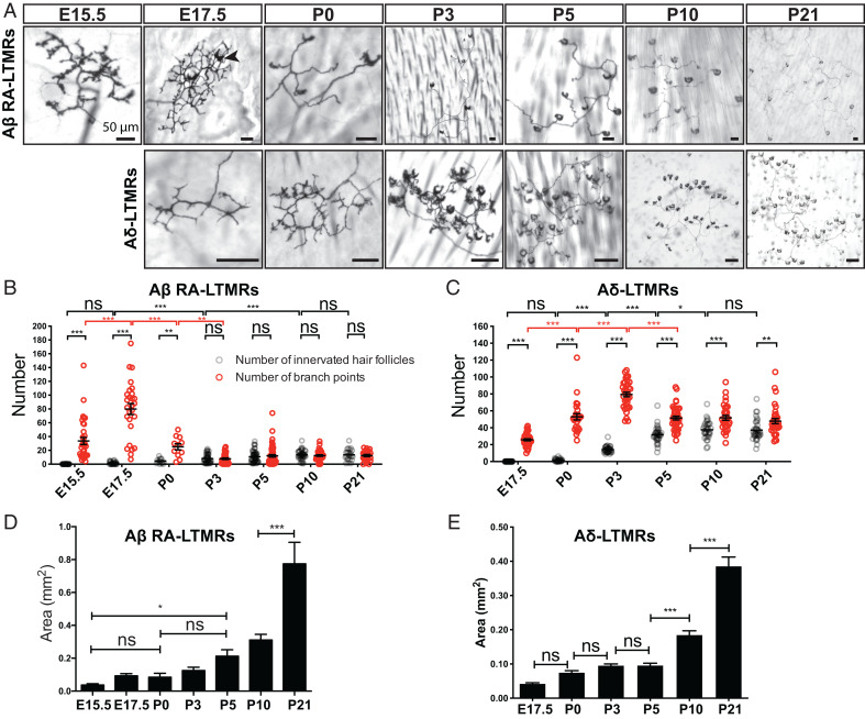
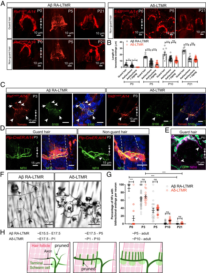
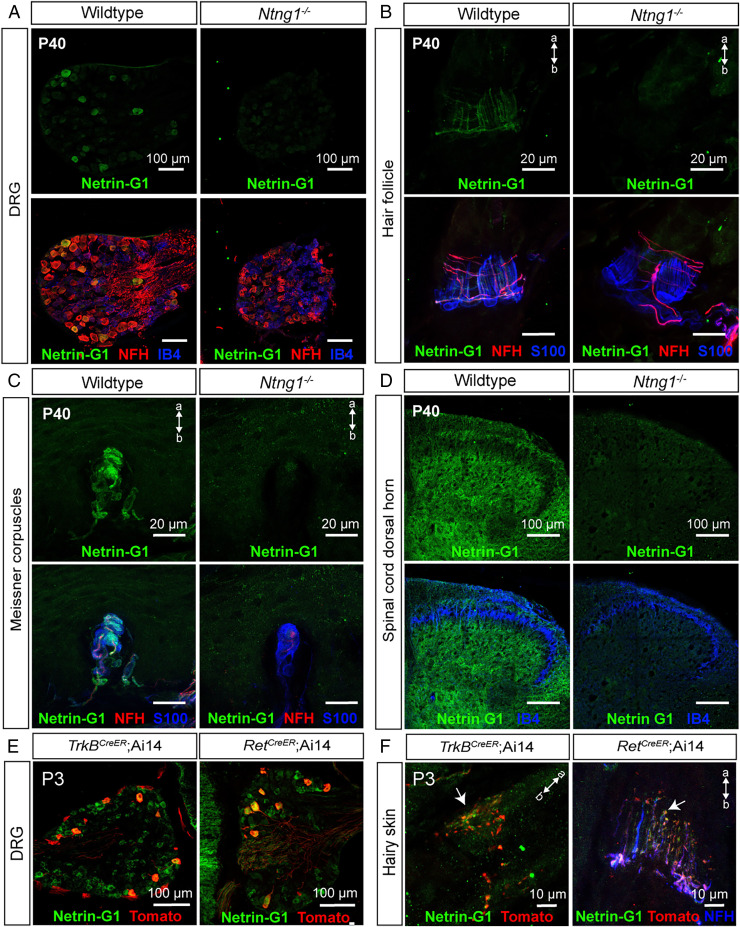
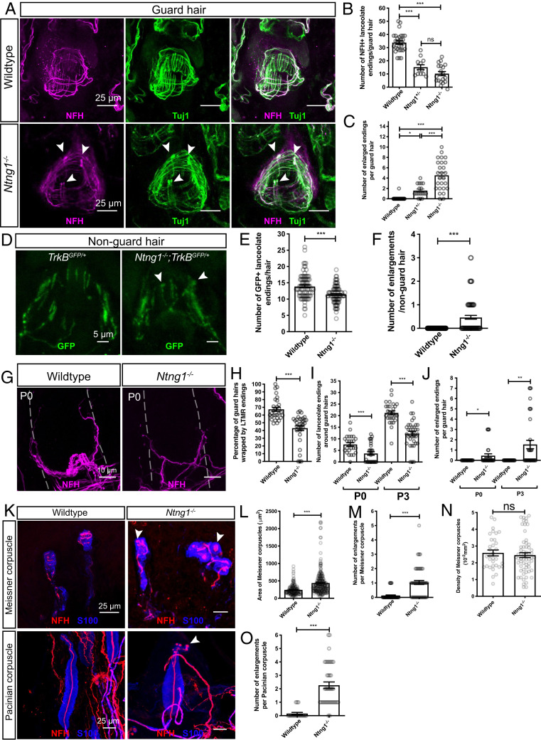
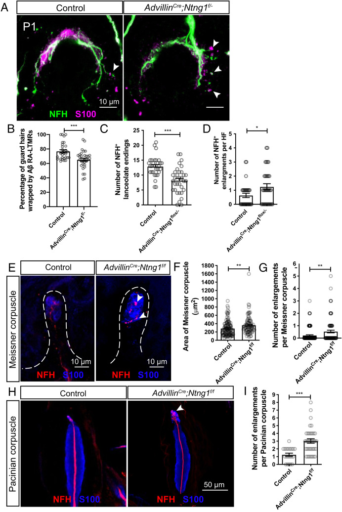
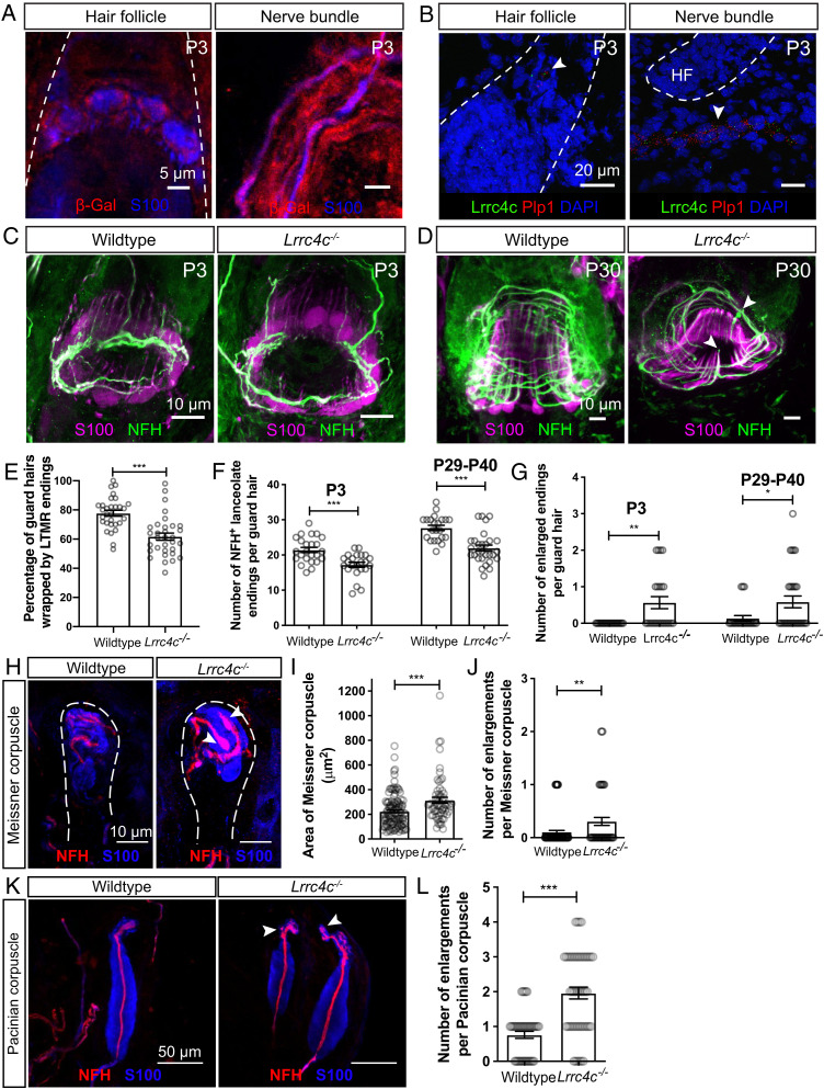
Low-threshold mechanoreceptors (LTMRs) and their cutaneous end organs convert light mechanical forces acting on the skin into electrical signals that propagate to the central nervous system. In mouse hairy skin, hair follicle-associated longitudinal lanceolate complexes, which are end organs comprising LTMR axonal endings that intimately associate with terminal Schwann cell (TSC) processes, mediate LTMR responses to hair deflection and skin indentation. Here, we characterized developmental steps leading to the formation of Aβ rapidly adapting (RA)-LTMR and Aδ-LTMR lanceolate complexes. During early postnatal development, Aβ RA-LTMRs and Aδ-LTMRs extend and prune cutaneous axonal branches in close association with nascent TSC processes. Netrin-G1 is expressed in these developing Aβ RA-LTMR and Aδ-LTMR lanceolate endings, and Ntng1 ablation experiments indicate that Netrin-G1 functions in sensory neurons to promote lanceolate ending elaboration around hair follicles. The Netrin-G ligand (NGL-1), encoded by Lrrc4c, is expressed in TSCs, and ablation of Lrrc4c partially phenocopied the lanceolate complex deficits observed in Ntng1 mutants. Moreover, NGL-1-Netrin-G1 signaling is a general mediator of LTMR end organ formation across diverse tissue types demonstrated by the fact that Aβ RA-LTMR endings associated with Meissner corpuscles and Pacinian corpuscles are also compromised in the Ntng1 and Lrrc4c mutant mice. Thus, axon-glia interactions, mediated in part by NGL-1-Netrin-G1 signaling, promote LTMR end organ formation.

Keywords: Netrin-G1; axon–glia interaction; development; lanceolate complex; pruning.


Conflict of interest statement

The authors declare no competing interest.

Figures

Fig. 1.
Fig. 1.
Peripheral arbors of Aβ RA-LTMRs and Aδ-LTMRs undergo pruning around birth. (A) Wholemount alkaline phosphatase staining of back hairy skin to visualize peripheral terminals of individual Aβ RA-LTMRs and Aδ-LTMRs at different developmental time points and at P21. The black arrowhead points to one example of a crescent-shaped axonal ending wrapped around a hair follicle. (Scale bars, 50 μm.) (B and C) Quantification of the number of innervated hair follicles (gray) and branch points (red) of individual Aβ RA-LTMRs (B, n = 257 neurons from 37 animals) and Aδ-LTMRs (C, n = 210 neurons from 17 animals). Each dot represents a single neuron. Two-way ANOVA with Tukey's multiple comparisons test and Šídák's multiple comparisons test. (D and E) Quantification of the area of skin covered by individual Aβ RA-LTMRs (D) and Aδ-LTMRs (E). One-way analysis of variance (ANOVA) with Šídák's multiple comparisons test. ns, not significant, *P < 0.05, **P < 0.01, ***P < 0.001.
Fig. 2.
Fig. 2.
Lanceolate endings of Aβ RA-LTMRs and Aδ-LTMRs emerge around hair follicles neonatally and closely associate with terminal Schwann cells. (A) Wholemount immunostaining images of back hairy skin showing Aβ RA-LTMRs in RetCreER; Ai14 animals and Aδ-LTMRs in TrkBCreER; Ai14 animals at P0, P5, and P21. Guard hairs were identified by TROMA-I (Krt8) staining that labels Merkel cells that assemble into touch domes. (B) Quantification of the length of lanceolate endings associated with hair follicles. Aβ RA-LTMRs (black) and Aδ-LTMRs (red) at P0, P5, P10, and P21, showing that Aβ RA-LTMR lanceolate endings innervating guard hairs develop earlier than the lanceolate endings innervating awl/auchene and zigzag hairs (together grouped as nonguard hairs). Aβ RA-LTMRs (n = 3 animals per time point): P0 (14 guard hairs and 28 nonguard hairs), P5 (14 guard hairs and 47 nonguard hairs), P10 (10 guard hairs and 39 nonguard hairs), and P21 (10 guard hairs and 15 nonguard hairs). Aδ-LTMRs (n = 3 animals per time point): P0 (8 nonguard hairs), P5 (32 nonguard hairs), P10 (49 nonguard hairs), and P21 (68 nonguard hairs). Each dot represents lanceolate ending length around a hair follicle. One-way ANOVA was used for each time point. (C) Back hairy skin sections from RetCreER; Ai14 animals (n = 3 animals) and TrkBCreER; Ai14 animals (n = 3 animals) were stained with an anti-Tomato antibody to label Aβ RA-LTMRs and Aδ-LTMR axonal terminals around hair follicles at P3. S100 (green) staining labels TSCs, and DAPI labels nuclei. White arrowheads point to some of the nascent lanceolate endings growing toward the apical and basal sides of the skin. White arrows and dotted lines denote cell bodies of TSCs. (D) Wholemount immunostaining images from Plp-CreER; Ai14 animals (back hairy skin, n = 3 animals), which were stained with an anti-Tomato antibody to label TSCs at P3. NFH (green) labels lanceolate endings of Aβ RA-LTMRs. White arrowhead points to nascent lanceolate endings growing toward the basal side of the skin. (E) Wholemount immunostaining image from a PLP-EGFP animal (back hairy skin, n = 3 animals), which was stained with an anti-GFP antibody to label TSCs at P0. White arrowhead points to the growing ends of the circumferential endings around the guard hair. (F) Wholemount alkaline phosphatase staining of back hairy skin reveals peripheral terminals from individual Aβ RA-LTMRs and Aδ-LTMRs sparsely labeled at P3. Insets showing a high-magnification region of lanceolate endings around hair follicles. Black arrowheads point to nascent lanceolate endings extending toward the basal side of the skin. (G) Quantification of the percentage of hair follicles with bidirectional endings per neuron for Aβ RA-LTMRs and Aδ-LTMRs at P0, P5, P10, and P21. Aβ RA-LTMRs: P0 (14 neurons from three animals), P3 (42 neurons from four animals), P5 (22 neurons from six animals), P10 (32 neurons from three animals), and P21 (21 neurons from three animals). Aδ-LTMRs: P0 (19 neurons from three animals), P3 (15 neurons from three animals), P5 (27 neurons from four animals), P10 (19 neurons from three animals), and P21 (27 neurons from three animals). HF, hair follicle. Each dot represents the percentage of hair follicles with bidirectional endings measured for a single neuron. Two-way ANOVA with Tukey's multiple comparisons test and Šídák's multiple comparisons test. (H) Summary of peripheral innervation steps for Aβ RA-LTMRs and Aδ-LTMRs. Black represents sensory axons, green cells represent terminal Schwann cells, and hair follicles are depicted in red. Dotted lines show that some sensory neuron branches and lanceolate endings are pruned during development. a, apical; b, basal. ns, not significant, *P < 0.05, **P < 0.01, ***P < 0.001.
Fig. 3.
Fig. 3.
Netrin-G1 is expressed in developing and adult Aβ RA-LTMRs and Aδ-LTMRs. (AD) Immunohistochemistry (IHC) images of T11–T13 DRG sections from wild-type and Ntng1−/− animals (n = 3 animals per genotype) at P40, showing expression of Netrin-G1 in NFH+ DRG neurons (A), lanceolate and circumferential endings around hair follicles (B), Aβ RA-LTMRs innervating Meissner corpuscles (C), and lumbar (L4) spinal cord (D). Netrin-G1 signal (green) is absent in Ntng1−/− animals. IB4 labels a large subset of nonpeptidergic sensory neurons and S100 labels TSCs. (E and F) Thoracic DRG and back hairy skin sections obtained from P3 RetCreER; Ai14 animals (n = 3 animals) and TrkBCreER; Ai14 animals (n = 3 animals) were stained with Netrin-G1 and Tomato to visualize Aβ RA-LTMRs and Aδ-LTMRs. NFH (blue) labels lanceolate endings of Aβ RA-LTMRs. a, apical; b, basal.
Fig. 4.
Fig. 4.
Netrin-G1 regulates the formation of lanceolate endings, Meissner corpuscles, and Pacinian corpuscles. (A) Wholemount immunostaining images of guard hairs in back hairy skin of adult wild-type and Ntng1−/− animals. Aβ RA-LTMRs lanceolate endings are marked by NFH (magenta) and Tuj1 (green) labeling. (B and C) Quantification of the number of NFH+ lanceolate endings (B) and the number of enlarged endings (C) per guard hair for Aβ RA-LTMRs in wild-type (B: 29 hair follicles from five animals; C: 42 hair follicles from seven animals), Ntng1+/− (B: 12 hair follicles from two animals; C: 17 hair follicles from two animals) and Ntng1−/− (B: 19 hair follicles from three animals; C: 26 hair follicles from three animals) animals, showing fewer Aβ RA-LTMR lanceolate endings in Ntng1+/− and Ntng1−/− animals. Each dot represents a single hair follicle. One-way ANOVA test. (D) Wholemount immunostaining images of nonguard hairs in back hairy skin in TrkBGFP/+ and Ntng1−/−; TrkBGFP/+ animals. Aδ-LTMRs lanceolate endings are marked by GFP labeling. (E and F) Quantification of the number of GFP+ lanceolate endings (E) and the number of enlarged endings (F) in TrkBGFP/+ (90 hair follicles from three animals) and Ntng1−/−; TrkBGFP/+ (95 hair follicles from three animals) animals, showing similar deficits in lanceolate ending formation with nonguard hairs. Student’s unpaired t test. (G) Wholemount immunostaining images of guard hairs in back hairy skin of P0 wild-type and Ntng1−/− animals. (H) Quantification of the percentages of guard hairs wrapped by NFH+ lanceolate endings at P0 in wild-type (29 hair follicles from three animals) and Ntng1−/− (33 hair follicles from three animals) animals. Student’s unpaired t test. (I and J) Quantification of the number of NFH+ lanceolate endings (I) and the number of enlarged endings (J) per guard hair in back hairy skin for Aβ RA-LTMRs in wild-type (25 hair follicles from three animals for P0; 28 hair follicles from five animals for P3) and Ntng1−/− (31 hair follicles from three animals for P0; 25 hair follicles from five animals for P3) animals. Student’s unpaired t test. (K) Representative IHC images of forepaw glabrous skin sections and Pacinian corpuscles. Meissner corpuscles and Pacinian corpuscles are labeled by S100 (blue) for visualizing lamellar cells and NFH (red) for visualizing Aβ RA-LTMRs. Arrowheads point to axonal enlargements. (LN) Quantification of the area (L), number of enlargements (M), and density (N) of Meissner corpuscles in the epidermis of wild-type (44 skin sections from four animals) and Ntng1−/− (60 skin sections from four animals) mice. Each dot represents a single skin section (O). Student’s unpaired t test. (O) Quantification of the number of enlargements per Pacinian corpuscle from wild-type (19 corpuscles from three animals) and Ntng1−/− (41 corpuscles from three animals) mice. Student’s unpaired t test. Each dot represents a single Pacinian corpuscle. ns, not significant, *P < 0.05, **P < 0.01, ***P < 0.001.
Fig. 5.
Fig. 5.
Netrin-G1 functions in somatosensory neurons to regulate mechanosensory end organ formation. (A) Wholemount IHC images of guard hairs in back hairy skin of P1 control (Ntng1+/−) and AdvillinCre; Ntng1f/− animals. (B) Quantification of the percentage of guard hairs wrapped by NFH+ lanceolate endings of P1 control (30 hair follicles from three animals) and AdvillinCre; Ntng1f/− (33 hair follicles from three animals) animals. (C and D) Quantification of the number of NFH+ lanceolate endings (C) and the number of enlarged endings (D) per guard hair in P1 control (30 hair follicles from three animals) and AdvillinCre; Ntng1f/− (33 hair follicles from three animals) mice. (E) Representative IHC images of forepaw glabrous skin sections, showing Meissner corpuscles in control and AdvillinCre; Ntng1f/f mice. Arrowheads point to axonal enlargements. (F and G) Quantification of the area (F) and number of enlargements (G) of Meissner corpuscles in the epidermis of control (32 sections from three animals) and AdvillinCre; Ntng1f/f (31 sections from three animals) mice. (H and I) Representative IHC images of Pacinian corpuscles (H) and quantification of the number of enlargements per Pacinian corpuscle (I) in littermate control (20 corpuscles from two animals) and AdvillinCre; Ntng1f/f (36 corpuscles from three animals) mice. The arrowhead points to axonal enlargements. Each dot represents a single Pacinian corpuscle. Student’s unpaired t test. *P < 0.05, **P < 0.01, ***P < 0.001.
Fig. 6.
Fig. 6.
NGL-1 is expressed in terminal Schwann cells and contributes to the formation of lanceolate endings, Meissner corpuscles, and Pacinian corpuscles. (A) Wholemount IHC images of P3 back hairy skin sections (n = 3 animals), showing that β-Gal (red) is detected in S100+ terminal Schwann cells and nonmyelinating Schwann cells (blue). Hair follicles are outlined with white dotted lines. Skin from wild-type animals was used as negative controls and no β-Gal signal was observed. (B) Example images of smRNA-FISH for P3 back hairy skin sections (n = 3 animals). White arrowheads denote the detection of Lrrc4c mRNA (green) near Plp1 mRNA (red). Hair follicles are outlined with white dotted lines. DAPI staining labels nuclei. (C and D) Wholemount immunostaining images of guard hairs in back hairy skin of P3 (C) and P30 (D) wild-type and Lrrc4c−/− animals. (E) Quantification of the percentage of guard hairs wrapped by NFH+ lanceolate endings in P0 wild-type (30 hair follicles from three animals) and Lrrc4c−/− animals (32 hair follicles from three animals). Each dot represents the result for one guard hair in back hairy skin. (F and G) Quantification of the number of NFH+ lanceolate endings (F) and the number of enlarged endings (G) in wild-type (24 hair follicles from four animals for P3; 22 hair follicles from three animals for P29–P40), and Lrrc4c−/− (23 hair follicles from three animals for P3; 29 hair follicles from four animals for P29–P40) animals. (H) Representative IHC images of forepaw glabrous skin sections of wild-type and Lrrc4c−/− animals. (I and J) Quantification of the area (I) and number of enlargements (J) of Meissner corpuscles in the wild-type (34 skin sections from four animals) and Lrrc4c−/− mice (32 skin sections from four animals). (K and L) Representative IHC images (K) and quantification of the number of enlargements per Pacinian corpuscle (L) of adult wild-type (20 corpuscles from three animals) and Lrrc4c−/− mice (36 corpuscles from three animals). Each dot represents a single Pacinian corpuscle. Student’s unpaired t test. *P < 0.05, **P < 0.01, ***P < 0.001.

Similar articles

Cited by

References

    1. Abraira V. E., Ginty D. D., The sensory neurons of touch. Neuron 79, 618–639 (2013). - PMC - PubMed
    1. Zimmerman A., Bai L., Ginty D. D., The gentle touch receptors of mammalian skin. Science 346, 950–954 (2014). - PMC - PubMed
    1. Handler A., Ginty D. D., The mechanosensory neurons of touch and their mechanisms of activation. Nat. Rev. Neurosci. 22, 521–537 (2021). - PMC - PubMed
    1. Horch K. W., Tuckett R. P., Burgess P. R., A key to the classification of cutaneous mechanoreceptors. J. Invest. Dermatol. 69, 75–82 (1977). - PubMed
    1. Le Pichon C. E., Chesler A. T., The functional and anatomical dissection of somatosensory subpopulations using mouse genetics. Front. Neuroanat. 8, 21 (2014). - PMC - PubMed

Publication types