Impairments of retinal hemodynamics and oxygen metrics in ocular hypertension-induced ischemia-reperfusion
- PMID: 36252653
- PMCID: PMC10985794
- DOI: 10.1016/j.exer.2022.109278
Impairments of retinal hemodynamics and oxygen metrics in ocular hypertension-induced ischemia-reperfusion
Abstract
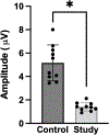
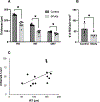
Ischemia-reperfusion (I/R) is an established model for retinal neurodegeneration. However, there is limited knowledge of retinal physiological metrics and their relationships to retinal function and morphology in the I/R model. The purpose of the study was to test the hypotheses that retinal hemodynamic and oxygen metrics are impaired and associated with visual dysfunction, retinal thinning, and retinal ganglion cell (RGC) loss due to I/R injury. Intraocular pressure (IOP) was increased in one eye of 10 rats for 90 min followed by reperfusion. Fellow eyes served as controls. After one week of reperfusion, multimodal imaging was performed to quantify total retinal blood flow (TRBF) and retinal vascular oxygen contents. Retinal oxygen delivery (DO2) and metabolism (MO2) were calculated. Pattern-evoked electroretinography (PERG) and optical coherence tomography were performed to measure RGC function and retinal thicknesses, respectively. RGCs were counted from retina whole mounts. After one week of reperfusion, TRBF was lower in study eyes than in control eyes (p < 0.0003). Similarly, DO2 and MO2 were reduced in study eyes compared to control eyes (p < 0.003). PERG amplitude, TRT, IRT, ORT, and RGCs were also lower in study eyes (p ≤ 0.01). DO2 and MO2 were correlated with PERG amplitude, TRT, IRT, and ORT (r ≥ 0.6, p ≤ 0.005). The findings improve knowledge of physiological metrics affected by I/R injury and have the potential for identifying biomarkers of injury and outcomes for evaluating experimental treatments.
Keywords: Blood flow; Ischemia-reperfusion; Oxygen delivery; Oxygen metabolism; Retinal thickness; Visual function.
Copyright © 2022 Elsevier Ltd. All rights reserved.
Conflict of interest statement
Declaration of Conflicting Interest
MS holds a patent for oxygen imaging technology. All authors have no conflict of interest.
Figures


References
-
- Adachi M, Takahashi K, Nishikawa M, Miki H, Uyama M, 1996. High intraocular pressure-induced ischemia and reperfusion injury in the optic nerve and retina in rats. Graefes Arch Clin Exp Ophthalmol 234, 445–451. - PubMed
-
- Büchi ER, Suivaizdis I, Fu J, 1991. Pressure-induced retinal ischemia in rats: an experimental model for quantitative study. Ophthalmologica 203, 138–147. - PubMed
Publication types
MeSH terms
Substances
Grants and funding
LinkOut - more resources
Full Text Sources
Medical

