

Performance Evaluation of the Schistoscope 5.0 for (Semi-)automated Digital Detection and Quantification of Schistosoma haematobium Eggs in Urine: A Field-based Study in Nigeria
- PMID: 36252803
- PMCID: PMC9709021
- DOI: 10.4269/ajtmh.22-0276
Performance Evaluation of the Schistoscope 5.0 for (Semi-)automated Digital Detection and Quantification of Schistosoma haematobium Eggs in Urine: A Field-based Study in Nigeria
Abstract
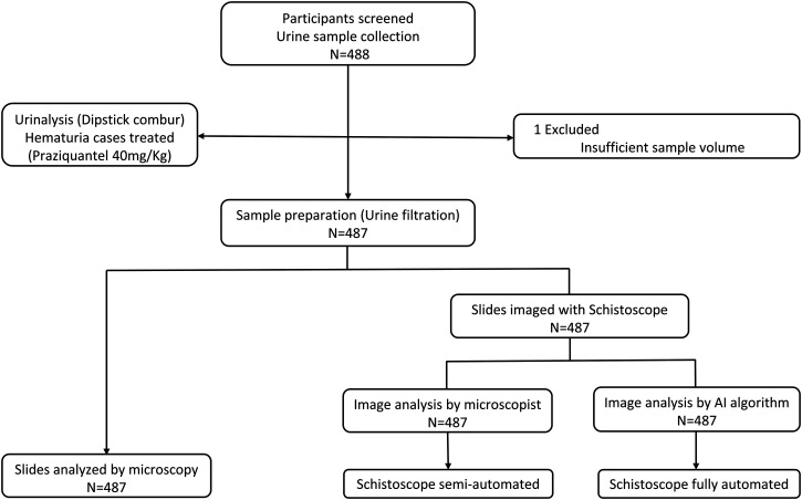
Conventional microscopy is the standard procedure for the diagnosis of schistosomiasis, despite its limited sensitivity, reliance on skilled personnel, and the fact that it is error prone. Here, we report the performance of the innovative (semi-)automated Schistoscope 5.0 for optical digital detection and quantification of Schistosoma haematobium eggs in urine, using conventional microscopy as the reference standard. At baseline, 487 participants in a rural setting in Nigeria were assessed, of which 166 (34.1%) tested S. haematobium positive by conventional microscopy. Captured images from the Schistoscope 5.0 were analyzed manually (semiautomation) and by an artificial intelligence (AI) algorithm (full automation). Semi- and fully automated digital microscopy showed comparable sensitivities of 80.1% (95% confidence interval [CI]: 73.2-86.0) and 87.3% (95% CI: 81.3-92.0), but a significant difference in specificity of 95.3% (95% CI: 92.4-97.4) and 48.9% (95% CI: 43.3-55.0), respectively. Overall, estimated egg counts of semi- and fully automated digital microscopy correlated significantly with the egg counts of conventional microscopy (r = 0.90 and r = 0.80, respectively, P < 0.001), although the fully automated procedure generally underestimated the higher egg counts. In 38 egg positive cases, an additional urine sample was examined 10 days after praziquantel treatment, showing a similar cure rate and egg reduction rate when comparing conventional microscopy with semiautomated digital microscopy. In this first extensive field evaluation, we found the semiautomated Schistoscope 5.0 to be a promising tool for the detection and monitoring of S. haematobium infection, although further improvement of the AI algorithm for full automation is required.
Figures


References
-
- World Health Organization , 2021. Schistosomiasis and soil-transmitted helminthiases: treating millions of people, despite the pandemic. Available at: https://www.who.int/news/item/08-12-2021-schistosomiasis-and-soil-transm.... Accessed January 27, 2022.
-
- McManus DP, Dunne DW, Sacko M, Utzinger J, Vennervald BJ, Zhou XN, 2018. Schistosomiasis. Nat Rev Dis Primers 4: 13. - PubMed
-
- Bustinduy AL, Randriansolo B, Sturt AS, Kayuni SA, Leustcher PDC, Webster BL, Van Lieshout L, Stothard JR, Feldmeier H, Gyapong M, 2022. An update on female and male genital schistosomiasis and a call to integrate efforts to escalate diagnosis, treatment and awareness in endemic and non-endemic settings: the time is now. Adv Parasitol 115: 1–44. - PubMed
-
- World Health Organization , 2020. Ending the Neglect to Attain the Sustainable Development Goals—A Road Map for Neglected Tropical Diseases 2021–2030. Geneva, Switzerland: WHO; 2020. License: CC BY-NC-SA 3.0 IGO.
MeSH terms
Substances
LinkOut - more resources
Full Text Sources
