. 2022 Nov;9(33):e2203257.
doi: 10.1002/advs.202203257. Epub 2022 Oct 17.

Nanoneedles Induce Targeted siRNA Silencing of p16 in the Human Corneal Endothelium

Affiliations

Nanoneedles Induce Targeted siRNA Silencing of p16 in the Human Corneal Endothelium

Eleonora Maurizi et al. Adv Sci (Weinh). 2022 Nov.

Abstract

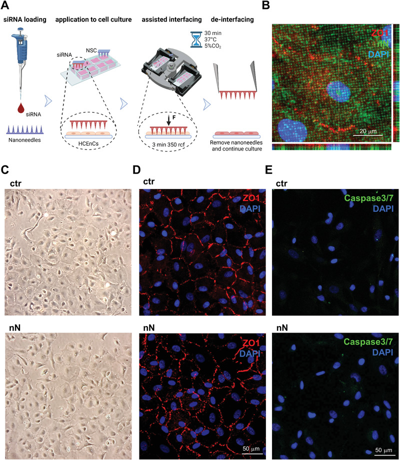
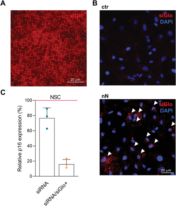
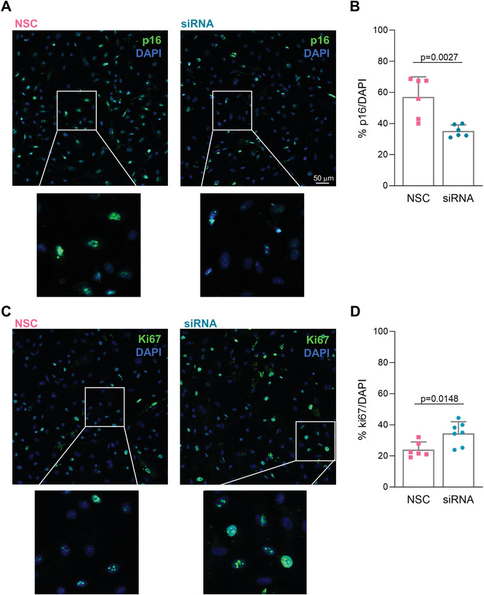
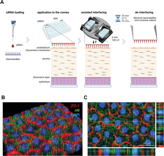
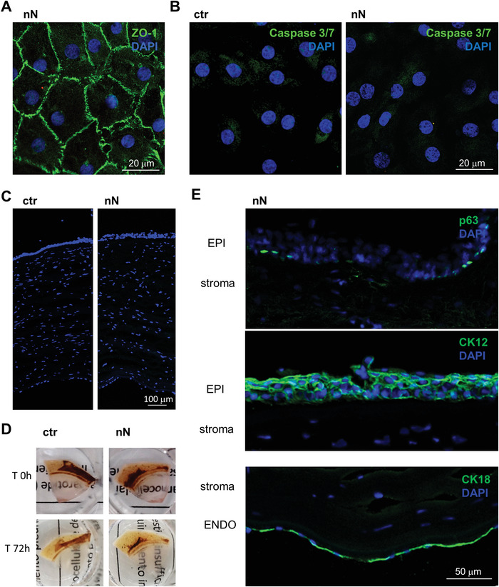
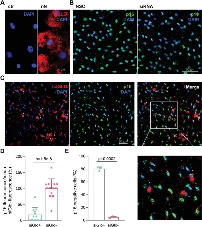
Nanoneedles can target nucleic acid transfection to primary cells at tissue interfaces with high efficiency and minimal perturbation. The corneal endothelium is an ideal target for nanoneedle-mediated RNA interference therapy aimed at enhancing its proliferative capacity, necessary for tissue regeneration. This work develops a strategy for siRNA nanoninjection to the human corneal endothelium. Nanoneedles can deliver p16-targeting siRNA to primary human corneal endothelial cells in vitro without toxicity. The nanoinjection of siRNA induces p16 silencing and increases cell proliferation, as monitored by ki67 expression. Furthermore, siRNA nanoinjection targeting the human corneal endothelium is nontoxic ex vivo, and silences p16 in transfected cells. These data indicate that nanoinjection can support targeted RNA interference therapy for the treatment of endothelial corneal dysfunction.

Keywords: gene therapy; nanoneedles; porous silicon; regenerative medicine; siRNA.


Conflict of interest statement

The authors declare no conflict of interest.

Figures

Figure 1
Figure 1
Nanoneedle interfacing with human corneal endothelial cells in vitro. A) Schematic representation of the nanoinjection approach for cultured primary HCEnCs. Image created with Biorender.com. B) Confocal microscopy orthogonal projections of nanoneedles (FITC labeled, green) interfaced with the cytosol, outlined by ZO‐1, and the nucleus of HCEnCs. Nanoneedles colocalize with HCEnCs. ZO‐1 staining (red) with DAPI (blue) nuclear counterstain. Scale bar 20 µm. Images were obtained immediately after nanoneedle assisted interfacing by centrifugation. C) Phase‐contrast image of the primary HCEnCs culture showing retained morphology following nanoneedle interfacing (nN), similar to the untreated control (ctr). Images were obtained immediately after the deinterfacing. D) Immunofluorescence microscopy of HCEnCs showing retained hexagonal morphology and ZO‐1 marker upon nN interfacing (nN) as well as in untreated HCEnCs (ctr). ZO‐1 staining (red) with DAPI (blue) nuclear counterstain. Scale bar 50 µm. Images were obtained 72 h following nanoneedles interfacing. E) Immunofluorescence microscopy of Caspase 3/7 activation. Lack of nuclear staining with faint cytoplasmic staining 72 h following nanoneedle interfacing (nN), comparable to untreated control (ctr) demonstrate lack of Caspase 3/7 activation, indicating absence of apoptotic events. Caspase 3/7 (green) staining with DAPI (blue) nuclear counterstain. Scale bar 50 µm.
Figure 2
Figure 2
In vitro nanoinjection of p16 (CDKN2A) siRNA in HCEnCs. A) Fluorescence microscopy of nanoneedles loaded with siGlo siRNA. siRNA is adsorbed uniformly across the nanoneedles. Scale bar 20 µm. B) Fluorescence microscopy of HCEnCs 48 h following nanoinjection of siGlo. siRNA accumulates in the cytosol of the cells upon nanoinjection (nN), as compared with the untreated HCEnCs (ctr). White arrows indicate some of the highly transfected cells. siGlo signal (red) with DAPI (blue) nuclear counterstain. Scale bar 50 µm. C) RT‐PCR of p16 expression showing silencing 48 h following p16‐siRNA nanoinjection, normalized and compared to NSC (nonspecific control, pink line). Experiment performed on three primary HCEnCs strains derived from different donors at passage 1 in culture (n = 3). The bar on the left (dark blue) indicates overall silencing level, the bar on the right (light blue) is normalized to the fraction of siGlo‐transfected cells in culture. Data are expressed as mean + SD.
Figure 3
Figure 3
Effects of in vitro nanoinjection. A,B) Nanoinjection with p16‐siRNA (siRNA) induces p16 protein knockdown if compared to NSC nanoinjection (NSC) 72 h following interfacing. A) Immunofluorescence microscopy showing p16 protein expression. p16 staining (green) with DAPI (blue) nuclear counterstain. Scale bar 50 µm. B) Quantification of the fraction of cells expressing p16 for p16‐siRNA (siRNA) and NSC nanoinjected HCEnCs (n = 3). p16‐siRNA nanoinjected samples have a statistically significant lower fraction of p16‐positive cells. Data are expressed as mean + SD. Two‐sided t‐test was used to assess statistical significance, p = 0.0027. C,D) RNAi nanoinjection to HCEnCs enhances their proliferative capacity 72 h following interfacing. C) Immunofluorescence microscopy showing ki67 protein expression in p16‐siRNA (siRNA) and NSC treated HCEnCs in vitro. ki67 staining (green) with DAPI (blue) as nuclear counterstain. D) Quantification of the fraction of cells expressing ki67 protein in p16‐siRNA (siRNA) and NSC treated HCEnCs in vitro (n = 3). p16‐siRNA nanoinjected samples have a statistically significant higher fraction of ki67‐positive cells. Data are expressed as mean + SD. Two‐sided t‐test was used to assess statistical significance, p = 0.0148.
Figure 4
Figure 4
Nanoneedle interfacing with the explanted human corneal endothelium. A) Schematic representation of the nanoinjection approach for explanted human corneas. Image created with Biorender.com. B,C) Immunofluorescence confocal microscopy of the interface between nanoneedles and the endothelium of human cornea explants. Images were obtained immediately after nanoneedle assisted interfacing by centrifugation. Nanoneedles colocalized with HCEnCs and did not protrude beyond them. ZO‐1 (red) localizes in HCEnCs membrane, FITC (green) labels nN and DAPI (blue) nuclear counterstain. B) 3D reconstruction from Z‐stack. C) Orthogonal projections. Scale bar 20 µm.
Figure 5
Figure 5
Nanoneedle interfacing preserves cornea integrity. A) Immunofluorescence confocal microscopy image of the explanted corneal endothelium obtained 72 h after nanoneedles interfacing, showing a maintained native endothelial morphology. ZO‐1 (green) staining with DAPI (blue) nuclear counterstain. Scale bar 20 µm. B) Immunofluorescence microscopy of Caspase 3/7 activation 72 h after nanoneedles interfacing. Lack of nuclear staining with faint cytoplasmic staining, comparable to untreated control demonstrate lack of Caspase 3/7 activation, indicating absence of apoptotic events. Caspase 3/7 (green) staining with DAPI (blue) nuclear counterstain. Scale bar 20 µm. C) Fluorescence microscopy of ctr and nN treated corneas (10 µm OCT sections) stained with DAPI (blue) shows that corneal thickness was unchanged in either of the two conditions. Scale bar 100 µm. D) Representative images of explanted corneas before and after (72 h) nanoinjection, showing how corneal clarity is maintained. E) Fluorescence microscopy of nN treated corneal sections, showing the expected expression and localization of epithelial (EPI, p63, and CK12) and endothelial (ENDO, CK18) cell markers (green). DAPI (blue) counterstains nuclei. Scale bar 50 µm.
Figure 6
Figure 6
Effects of p16‐siRNA nanoinjection to the explanted human corneal endothelium. A) Immunofluorescence microscopy of siGlo nanoinjection to the endothelium of explanted human corneas. Images were obtained 48 h after nanoinjection. HCEnCs cells display cytosolic siGlO in the area of nanoinjection (nN) as compared to untreated controls (ctr). siGlo signal (red) with DAPI (blue) nuclear counterstain. Scale bar 20 µm. B) Immunofluorescence microscopy of p16 protein expression in NSC and siRNA treated HCEnCs of explanted corneas. p16 (green) staining with DAPI (blue) nuclear counterstain. Scale bar 50 µm. C) Immunofluorescence microscopy of explanted human corneas 72 h following nanoinjection of p16 siRNA (n = 3). A significant correlation is visible between siGlo signal and loss of p16 signal, as highlighted by the white asterisks. p16 (green), siGlo (red) staining with DAPI (blue) nuclear counterstain. Scale bar 50 µm. D) Immunofluorescence quantification evaluating the fraction of p16 negative cells in siGlo+ transfected and siGlo‐ untransfected HCEnCs. Values are represented as mean + SD. Two‐sided t‐test was used to assess statistical significance, p = 1.5e‐8. E) Immunofluorescence quantification of p16 expression levels in siGlo+ transfected and siGlo‐untransfected cells. Values are represented as mean + SD. Two‐sided t‐test was used to assess statistical significance, p = 0.0002.

References

    1. Joyce N. C., Navon S. E., Roy S., Zieske J. D., Invest. Ophthalmol. Visual Sci. 1996, 37, 1566. - PubMed
    1. Gain P., Jullienne R., He Z., Aldossary M., Acquart S., Cognasse F., Thuret G., JAMA Ophthalmol. 2016, 134, 167. - PubMed
    1. Van Den Bogerd B., Dhubhghaill S. N.­, Koppen C., Tassignon M.‐J. C., Zakaria N., Surv. Ophthalmol. 2018, 63, 149. - PubMed
    1. Català P., Thuret G., Skottman H., Mehta J. S., Parekh M., Dhubhghailll S. N., Collin R. W. J., Nuijts R. M. M. A., Ferrari S., LaPointe V. L. S., Dickman M. M., Prog. Retinal Eye Res. 2021, 87, 100987. - PubMed
    1. Kampik D., Ali R. R., Larkin D. F., Exp. Eye Res. 2012, 95, 54. - PubMed

Publication types

MeSH terms

Substances

LinkOut - more resources