. 2022 Nov 8;40(47):6839-6848.
doi: 10.1016/j.vaccine.2022.09.057. Epub 2022 Sep 26.

Characterization and immunogenicity of SARS-CoV-2 spike proteins with varied glycosylation

Affiliations

Characterization and immunogenicity of SARS-CoV-2 spike proteins with varied glycosylation

Tingting Deng et al. Vaccine. .

Abstract

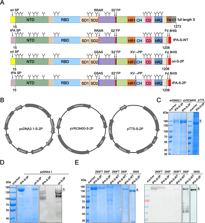
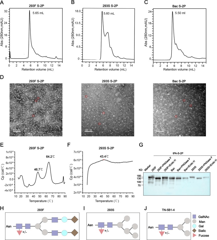
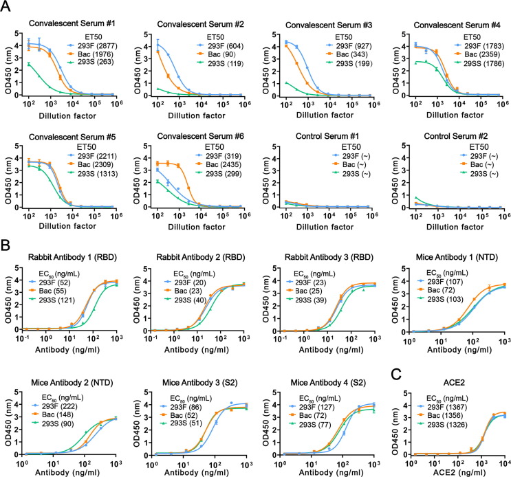
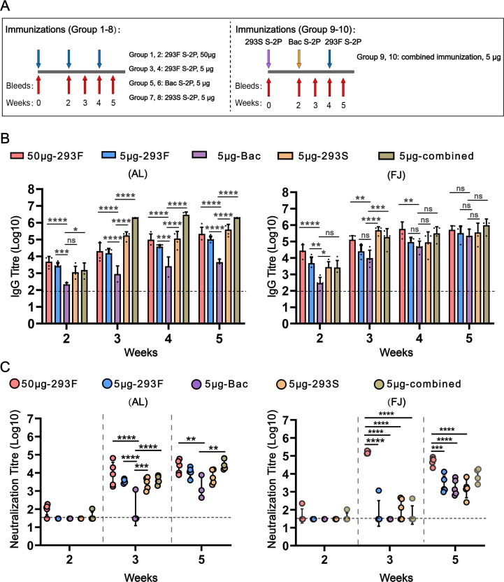
The ongoing coronavirus disease-19 (COVID-19) pandemic, caused by severe acute respiratory syndrome coronavirus 2 (SARS-CoV-2), has drastically changed our way of life and continues to have an unmitigated socioeconomic impact across the globe. Research into potential vaccine design and production is focused on the spike (S) protein of the virus, which is critical for virus entry into host cells. Yet, whether the degree of glycosylation in the S protein is associated with vaccine efficacy remains unclear. Here, we first optimized the expression of the S protein in mammalian cells. While we found no significant discrepancy in purity, homogeneity, or receptor binding ability among S proteins derived from 293F cells (referred to as 293F S-2P), 293S GnTI- cells (defective in N-acetylglucosaminyl transferase I enzyme; 293S S-2P), or TN-5B1-4 insect cells (Bac S-2P), there was significant variation in the glycosylation patterns and thermal stability of the proteins. Compared with the partially glycosylated 293S S-2P or Bac S-2P, the fully glycosylated 293F S-2P exhibited higher binding reactivity to convalescent sera. In addition, 293F S-2P induced higher IgG and neutralizing antibody titres than 293S or Bac S-2P in mice. Furthermore, a prime-boost-boost regimen, using a combined immunization of S-2P proteins with various degrees of glycosylation, elicited a more robust neutralizing antibody response than a single S-2P alone. Collectively, this study provides insight into ways to design a more effective SARS-CoV-2 immunogen.

Keywords: 293 cells; Glycosylation; Immunogenicity; SARS-CoV-2; Spike.


Conflict of interest statement

Declaration of Competing Interest The authors declare that they have no known competing financial interests or personal relationships that could have appeared to influence the work reported in this paper.

Figures

Fig. 1
Fig. 1
Construct design and expression optimization of the SARS CoV-2 Spike (S) protein. (A) Linear representation of the S protein primary structure and construct design. Full-length (FL) S protein, extracellular S protein with tPA signal peptide (tPA-S-WT) and extracellular S-2P protein with tPA (tPA-S-2P) or the original signal peptide (Ori-S-2P). NTD, N-terminal domain; RBD, receptor binding domain; SD1, subdomain 1, SD2, subdomain 2; FP Fusion peptide, HR1, heptad repeat 1, CH, central helix, CD, connector domain, HR2, heptad repeat 2; TM, transmembrane domain; tPA, tissue plasminogen activator; CT, cytoplasmic tail. (B) Construct map of pcDNA3.1, pVRC8400 and pTT5 vectors that were inserted with the tPA-S-2P gene at the multiple cloning site. (C) SDS-PAGE analysis of Ni-Sepharose excel-purified S-2P protein produced with different vectors. (D) SDS-PAGE and western blot analysis of purified S-2P from two constructs with different signal peptides. (E) SDS-PAGE and western blot analysis of purified tPA-S-2P and tPA-S-WT produced via three types of 293 cell lines. To make the western blot image more clearly, we down-adjusted the Gray coefficient of exposure parameter. Human convalescent serum was used for immunoblotting.
Fig. 2
Fig. 2
Characterization of purified SARS-CoV-2 spike (S) protein. (A-C) 293F S-2P, 293S S-2P, and Bac S-2P proteins were analysed by high-pressure size-exclusion chromatography (HPSEC). (D) Negative-stain EM micrographs of purified 293F S-2P, 293S S-2P, and Bac S-2P proteins. (E-F) Differential scanning calorimetry (DSC) profiles of 293F S-2P and 293S S-2P. Two thermal denaturation midpoints (Tm) are shown for 293F S-2P at 46.7℃ (Tm1) and 64.2℃ (Tm2), but just one weak peak at 43.4℃ for 293S S-2P. (G) Western blot of 293F-S-2P, 293S-S-2P, Bac-S-2P proteins following treatment with endo-H or PNGase F. Untreated samples act as the control. (H-J) The glycosylation anatomy of proteins produced by 293F cell, 293S GnTI- and Tn-5B1-4 cells (insect cell). Different glycan types are labelled with different colours and shapes.
Fig. 3
Fig. 3
Antigenicity and receptor binding of S-2P proteins. (A) Reactivities of S-2P proteins against COVID-19 convalescent human sera (#1-#6) and control sera (#1-#2), ∼ denotes not detected. (B) Reactivities of S-2P proteins against three rabbit monoclonal antibodies targeting the receptor-binding domain (RBD), two mouse monoclonal antibodies targeting the N-terminal domain (NTD) and two antibodies targeting the S2 subunit. (C) Reactivities of S-2P proteins to the ACE2 receptor.
Fig. 4
Fig. 4
Immunogenicity of SARS-CoV-2 spike (S) proteins. (A) Mice immunization schedules. Group 1, 3, 5, 7, 9 received immunizations formulated with aluminium adjuvant. Group 2, 4, 6, 8, 10 received immunizations formulated with Freund’s adjuvant. (B) Antigen-specific IgG antibody titres induced by ten immunized groups. (C) Neutralizing antibody titres against SARS-CoV-2 pseudovirus (Wuhan-Hu-1 strain). The dotted line indicates the limit of detection for the assay. Statistical analysis was performed by one-way ANOVA. Significant differences were determined using the Holm-Sidak pairwise multiple comparisons in Graphpad Prism 8.0 (ns = non-significant; *P < 0.1, **P < 0.01, ***P < 0.001, ****P < 0.0001). (A) Groups statistically different from the Bac S-2P group are indicated. (B) Groups statistically different from each other are indicated. An absence of symbols indicates no statistical difference between the groups.

References

    1. Petrosillo N., Viceconte G., Ergonul O., Ippolito G., Petersen E. COVID-19, SARS and MERS: are they closely related? Clin Microbiol Infect. 2020;26(6):729–734. - PMC - PubMed
    1. Zhuang Z., Lai X., Sun J., et al. Mapping and role of T cell response in SARS-CoV-2-infected mice. J Exp Med. 2021;218(4) - PMC - PubMed
    1. Hoffmann M., Arora P., Groß R., Seidel A., Hörnich B.F., Hahn A.S., et al. SARS-CoV-2 variants B.1.351 and P.1 escape from neutralizing antibodies. Cell. 2021;184(9):2384–2393.e12. - PMC - PubMed
    1. Li Q., Nie J., Wu J., Zhang L., Ding R., Wang H., et al. SARS-CoV-2 501Y.V2 variants lack higher infectivity but do have immune escape. Cell. 2021;184(9):2362–2371.e9. - PMC - PubMed
    1. Li Q., Wu J., Nie J., Zhang L., Hao H., Liu S., et al. The impact of mutations in SARS-CoV-2 spike on viral infectivity and antigenicity. Cell. 2020;182(5):1284–1294.e9. - PMC - PubMed

Publication types

Substances