. 2022 Oct 17;12(1):17373.
doi: 10.1038/s41598-022-20411-4.

A lightweight convolutional neural network model with receptive field block for C-shaped root canal detection in mandibular second molars

Affiliations

A lightweight convolutional neural network model with receptive field block for C-shaped root canal detection in mandibular second molars

Lijuan Zhang et al. Sci Rep. .

Abstract

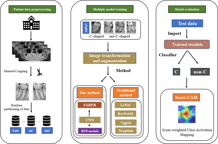
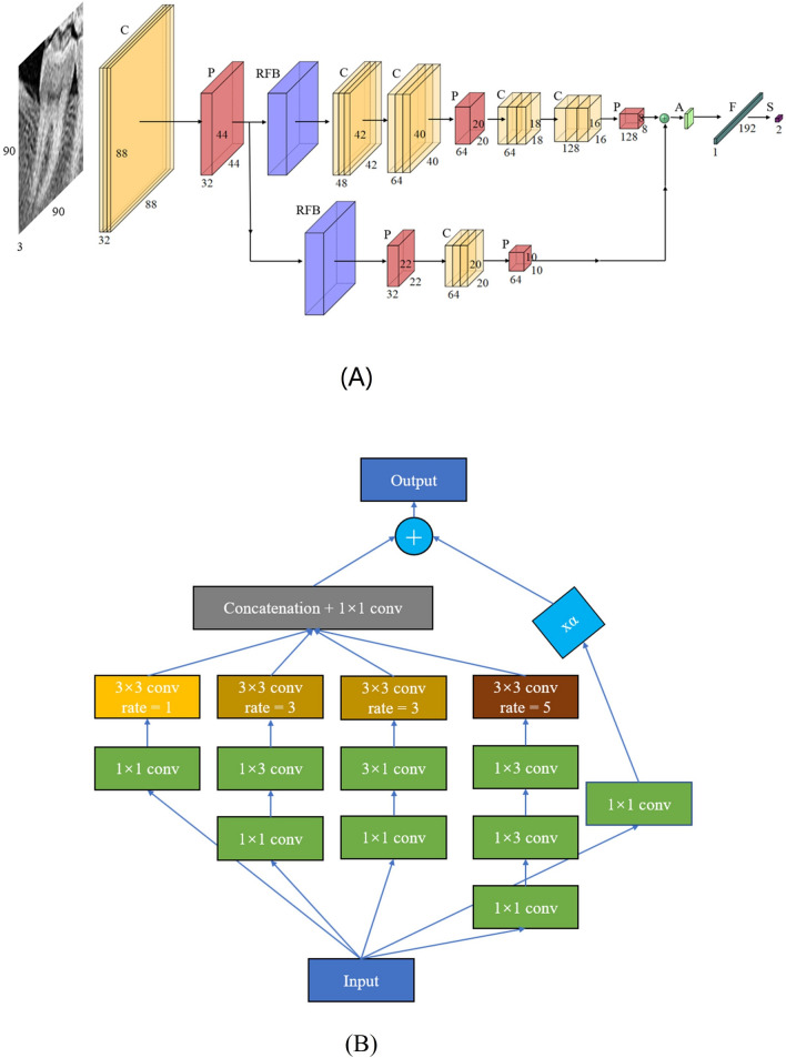
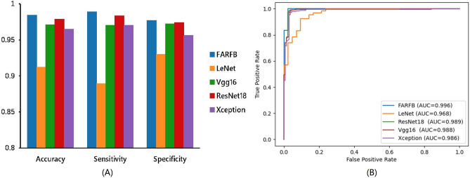
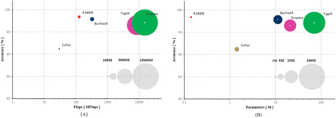
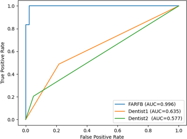
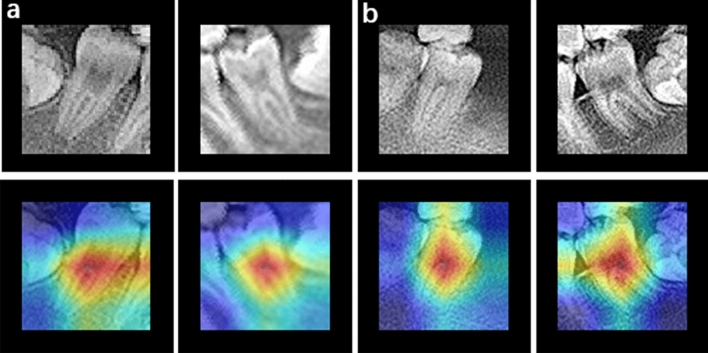
Rapid and accurate detection of a C-shaped root canal on mandibular second molars can assist dentists in diagnosis and treatment. Oral panoramic radiography is one of the most effective methods of determining the root canal of teeth. There are already some traditional methods based on deep learning to learn the characteristics of C-shaped root canal tooth images. However, previous studies have shown that the accuracy of detecting the C-shaped root canal still needs to be improved. And it is not suitable for implementing these network structures with limited hardware resources. In this paper, a new lightweight convolutional neural network is designed, which combined with receptive field block (RFB) for optimizing feature extraction. In order to optimize the hardware resource requirements of the model, a lightweight, multi-branch, convolutional neural network model was developed in this study. To improve the feature extraction ability of the model for C-shaped root canal tooth images, RFB has been merged with this model. RFB has achieved excellent results in target detection and classification. In the multiscale receptive field block, some small convolution kernels are used to replace the large convolution kernels, which allows the model to extract detailed features and reduce the computational complexity. Finally, the accuracy and area under receiver operating characteristics curve (AUC) values of C-shaped root canals on the image data of our mandibular second molars were 0.9838 and 0.996, respectively. The results show that the deep learning model proposed in this paper is more accurate and has lower computational complexity than many other similar studies. In addition, score-weighted class activation maps (Score-CAM) were generated to localize the internal structure that contributed to the predictions.


Conflict of interest statement

The authors declare no competing interests.

Figures

Figure 1
Figure 1
Evaluation of panoramic image datasets by two dentists and the Kappa test coefficient of the ground truth.
Figure 2
Figure 2
The image data flow chart defines how to decompose the data set.
Figure 3
Figure 3
The workflow chart for classification of the tooth root canal. Cut out the mandibular second molar tooth image from the panoramic image produced from the original image data. The tooth image data obtained in the previous step was inputted into different network systems to classify and compare the results. The tooth image data gained in the last step was inputted to the proposed network combining increasing receptive field modules and classical network structure method. Finally, Score-CAM is used to visualize where the network focuses when making classification decisions.
Figure 4
Figure 4
The network structure of FARFB (A). The capital letters A, C, F, P, S and S denote the Global Average Pooling, Convolutional, Full Connection, Pooling and SoftMax respectively. The values represent the number of channels, width and height of the feature maps. The composition structure of receptive field block (RFB) (B).
Figure 5
Figure 5
The details of the train set, validation set and test set composition.
Figure 6
Figure 6
Comparison of the proposed method with traditional methods in accuracy, sensitivity, specificity (A). Receiver operating characteristic (ROC) curves for the proposed method and traditional methods (B).
Figure 7
Figure 7
The graphical performance of each network model, the number of its parameters and calculations in C-shaped and non-C-shaped root canal classification tasks (the input image size of Vgg16 is 224 × 224. The input image size of Xception is 299 × 299). The gray circles are the standard display of the number of parameters or calculations at each level.
Figure 8
Figure 8
Receiver operating characteristic (ROC) curves for the proposed method and dentists.
Figure 9
Figure 9
Score-CAM output for C-shaped and non-C-shaped type fractures classification. C-shaped root canal tooth images (a) and non-C-shaped root canal tooth images (b). Original images (top) and overlapped on the original image (bottom). The visualization certifies that the network is focusing on the correct area of the tooth.

References

    1. Alfawaz H, et al. Prevalence of C-shaped canal system in mandibular first and second molars in a Saudi population assessed via cone beam computed tomography: A retrospective study. Clin. Oral Invest. 2019;23:107–112. doi: 10.1007/s00784-018-2415-0. - DOI - PubMed
    1. Cooke HG, 3rd, Cox FL. C-shaped canal configurations in mandibular molars. J. Am. Dent. Assoc. 1979;1939(99):836–839. doi: 10.14219/jada.archive.1979.0402. - DOI - PubMed
    1. Walker RT. Root form and canal anatomy of mandibular second molars in a Southern Chinese population. J. Endod. 1988;14:325–329. doi: 10.1016/S0099-2399(88)80192-4. - DOI - PubMed
    1. Solomonov M, Paqué F, Fan B, Eilat Y, Berman LH. The challenge of C-shaped canal systems: A comparative study of the self-adjusting file and ProTaper. J. Endod. 2012;38:209–214. doi: 10.1016/j.joen.2011.10.022. - DOI - PubMed
    1. Manning SA. Root canal anatomy of mandibular second molars: Part II C-shaped canals. Int. Endod. J. 1990;23:40–45. doi: 10.1111/j.1365-2591.1990.tb00801.x. - DOI - PubMed

Publication types

MeSH terms