. 2023 Jan;20(1):195-206.
doi: 10.1007/s13311-022-01308-6. Epub 2022 Oct 17.

Lecanemab, Aducanumab, and Gantenerumab - Binding Profiles to Different Forms of Amyloid-Beta Might Explain Efficacy and Side Effects in Clinical Trials for Alzheimer's Disease

Affiliations

Lecanemab, Aducanumab, and Gantenerumab - Binding Profiles to Different Forms of Amyloid-Beta Might Explain Efficacy and Side Effects in Clinical Trials for Alzheimer's Disease

Linda Söderberg et al. Neurotherapeutics. 2023 Jan.

Abstract

Immunotherapy against amyloid-beta (Aβ) is a promising option for the treatment of Alzheimer's disease (AD). Aβ exists as various species, including monomers, oligomers, protofibrils, and insoluble fibrils in plaques. Oligomers and protofibrils have been shown to be toxic, and removal of these aggregates might represent an effective treatment for AD. We have characterized the binding properties of lecanemab, aducanumab, and gantenerumab to different Aβ species with inhibition ELISA, immunodepletion, and surface plasmon resonance. All three antibodies bound monomers with low affinity. However, lecanemab and aducanumab had very weak binding to monomers, and gantenerumab somewhat stronger binding. Lecanemab was distinctive as it had tenfold stronger binding to protofibrils compared to fibrils. Aducanumab and gantenerumab preferred binding to fibrils over protofibrils. Our results show different binding profiles of lecanemab, aducanumab, and gantenerumab that may explain clinical results observed for these antibodies regarding both efficacy and side effects.

Keywords: Aducanumab; Amyloid-beta species; Gantenerumab; Lecanemab; Therapeutic antibodies.


Conflict of interest statement

LS, MJ, PN, HL, FE, GO, CM, and LL are employees and shareholders of BioArctic. LL is a co‐founder and board member of BioArctic.

Figures

Fig. 1
Fig. 1
Principle of the inhibition ELISA. Two steps are involved in the inhibition ELISA. Step one: incubation of Aβ species with the investigated antibodies for specific binding. Step two: the antibody-antigen mixtures from step one, containing complexes and free antibodies, are added to the plate coated with Aβ-protofibrils to allow binding of free antibody. Antigen binding in step one will then consequently reduce binding to the coated Aβ-protofibrils. As a result of prior binding of sample antigen to primary antibody, the reaction in the ELISA plate wells is reduced and the antigen concentration required to inhibit half of the maximum signal in the inhibition ELISA is defined as IC50, which was used as an estimate of the antibody’s affinity and selectivity for the investigated antigen
Fig. 2
Fig. 2
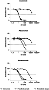
Results from inhibition ELISA with binding to monomeric Aβ and small and large protofibrils. Normalized and blank subtracted OD405 values were plotted against Aβ concentration. Curves represent mean ± SD for monomeric Aβ, squares, small protofibrils, black crosses, and large protofibrils, black triangles
Fig. 3
Fig. 3
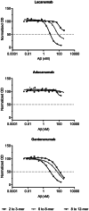
Results from the inhibition ELISA with small Aβ oligomers. Normalized and blank subtracted OD405 values were plotted against Aβ concentrations. Curves representing 8–12-mer, open circles, 6–8-mer, black triangles, and 2–3-mer, open squares
Fig. 4
Fig. 4
Evaluation of antibody binding to synthetic Aβ protofibrils in solution using immunodepletion. Antibodies, 0.67, 6.7, 67, 670, 6670 pM, were incubated with 10 pM of small (black columns) or large (white columns) Aβ protofibrils, followed by pull-down using magnetic protein A beads. Unbound Aβ protofibrils remained in the supernatant. Data expressed as % remaining protofibrils compared to bead control, and are presented as mean ± SD
Fig. 5
Fig. 5
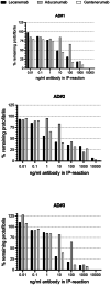
Immunodepletion of protofibrils from AD soluble brain extracts using lecanemab, aducanumab, and gantenerumab. Protofibrils remaining in the supernatant were analyzed using a protofibril specific assay (mAb158-1C3-bio). Data expressed as % remaining protofibril compared to bead control
Fig. 6
Fig. 6
Representative SPR sensorgrams (blue curves) and curve fittings (black curves) of binding to Aβ monomers, small and large protofibrils, and fibrils for lecanemab, aducanumab, and gantenerumab

References

    1. Glenner GG, Wong CW. Alzheimer’s disease and Down’s syndrome: sharing of a unique cerebrovascular amyloid fibril protein. Biochem Biophys Res Commun. 1984;122(3):1131–1135. doi: 10.1016/0006-291X(84)91209-9. - DOI - PubMed
    1. Masters CL, Simms G, Weinman NA, Multhaup G, McDonald BL, Beyreuther K. Amyloid plaque core protein in Alzheimer disease and Down syndrome. Proc Natl Acad Sci U S A. 1985;82(12):4245–4249. doi: 10.1073/pnas.82.12.4245. - DOI - PMC - PubMed
    1. Hardy J, Selkoe DJ. The amyloid hypothesis of Alzheimer’s disease: progress and problems on the road to therapeutics. Science. 2002;297(5580):353–356. doi: 10.1126/science.1072994. - DOI - PubMed
    1. Walsh DM, Lomakin A, Benedek GB, Condron MM, Teplow DB. Amyloid beta-protein fibrillogenesis. Detection of a protofibrillar intermediate. J Biol Chem. 1997;272(35):22364–72. - PubMed
    1. O'Nuallain B, Freir DB, Nicoll AJ, Risse E, Ferguson N, Herron CE, et al. Amyloid beta-protein dimers rapidly form stable synaptotoxic protofibrils. J Neurosci. 2010;30(43):14411–14419. doi: 10.1523/JNEUROSCI.3537-10.2010. - DOI - PMC - PubMed

MeSH terms