Review
. 2022 Nov;18(11):661-680.
doi: 10.1038/s41582-022-00723-9. Epub 2022 Oct 17.

Human cerebral organoids - a new tool for clinical neurology research

Affiliations
Review

Human cerebral organoids - a new tool for clinical neurology research

Oliver L Eichmüller et al. Nat Rev Neurol. 2022 Nov.

Abstract

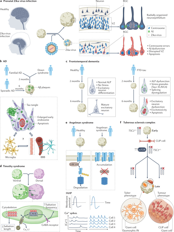
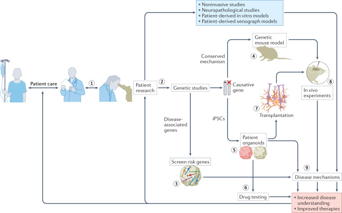
The current understanding of neurological diseases is derived mostly from direct analysis of patients and from animal models of disease. However, most patient studies do not capture the earliest stages of disease development and offer limited opportunities for experimental intervention, so rarely yield complete mechanistic insights. The use of animal models relies on evolutionary conservation of pathways involved in disease and is limited by an inability to recreate human-specific processes. In vitro models that are derived from human pluripotent stem cells cultured in 3D have emerged as a new model system that could bridge the gap between patient studies and animal models. In this Review, we summarize how such organoid models can complement classical approaches to accelerate neurological research. We describe our current understanding of neurodevelopment and how this process differs between humans and other animals, making human-derived models of disease essential. We discuss different methodologies for producing organoids and how organoids can be and have been used to model neurological disorders, including microcephaly, Zika virus infection, Alzheimer disease and other neurodegenerative disorders, and neurodevelopmental diseases, such as Timothy syndrome, Angelman syndrome and tuberous sclerosis. We also discuss the current limitations of organoid models and outline how organoids can be used to revolutionize research into the human brain and neurological diseases.


Conflict of interest statement

J.A.K. is on the supervisory and scientific advisory board of a:head bio AG (aheadbio.com) and is an inventor on several patents relating to cerebral organoids. O.L.E. declares no competing interests.

Figures

Fig. 1
Fig. 1. Organoids can bridge patient and animal studies to advance our understanding of neurological disease.
Studies in patients (left), such as sequencing, neuropathology or patient-derived xenograft models, provide a snapshot of disease at a given time point. Furthermore, these studies are usually not started until symptoms become apparent, meaning that the earliest pathogenic processes are not captured. Noninvasive and longitudinal studies to capture these early processes require large sample sizes and a lot of time. In animal studies (right), disease initiation can be controlled, so disease initiation, pathogenesis and progression can be studied throughout the disease course. Transfer of knowledge from animal studies to humans and vice versa relies on the assumption that disease mechanisms are conserved between humans and animal models, which is not always true. 3D human model systems such as organoids could be useful for bridging this gap, as they enable studies of early disease stages in human-derived tissue.
Fig. 2
Fig. 2. Innovations of human neurodevelopment.
The human brain develops over a protracted period of time (centre), resulting in its complex structure. This development involves several processes that are unique to humans (parts ae). a | Radial glial cell development. Apical radial glia cells (aRGCs, blue) are the neural stem cells that give rise to the human brain. aRGCs reside in the ventricular zone (VZ) and are connected to the ventricular surface via apical processes. At gestational week (GW) 14 (left), they pass through the cortical plate (CP) and connect to the pial surface via basal processes. Outer RGCs (oRGCs, brown) emerge in the subventricular zone (SVZ) and connect only to the pial surface via their basal process. Subsequently (at GW18, centre), the basal processes of aRGCs detach from the pial surface, and these cells become truncated radial glial cells (tRGCs). At this stage, the progenitor zone (right) is organized into a VZ that contains tRGCs, an inner SVZ (iSVZ) that contains intermediate progenitor cells (IPCs, dark yellow) and an outer SVZ (oSVZ) that contains oRGCs. Newly generated neurons (red) ascend along the basal processes of the radial glial cells towards the CP. b | Expansion of cortical layers II and III. Excitatory neurons are generated in an inside–out manner. Neurons migrate through the intermediate zone (IZ) towards the CP, which is delineated by the marginal zone (MZ) towards the pial surface. The first neurons to be generated are subplate neurons (dark blue), which form the subplate (SP). The deep SP and upper SP are formed sequentially. In humans, the SP expands greatly during development (compare GW13.5 with GW26–29) — during mid-gestation, the SP becomes larger than the CP and cortical layers I to VI combined. At later stages, an increased contribution of oRGCs to neurogenesis results in expansion of cortical layers II and III in the human brain (purple; compare GW26–29 with newborn). The SP also reduces in size and the prominent white matter (WM) emerges. c | Interneuron generation in the ventral forebrain within the ganglionic eminences. The human medial ganglionic eminence (MGE, left) contains doublecortin-positive cell-enriched nests (yellow) that contribute to neuronal production during the later stages of development. The MGE, lateral ganglionic eminence (LGE) and caudal ganglionic eminence (CGE) generate interneurons throughout neurogenesis (right) but the peak of neurogenesis in the CGE is later than in other regions and persists until the end of gestation. d | Interneuron migration to the cortex. In humans, this process persists until the first years of life, with large corridors of interneurons migrating in the so-called Arc into the forebrain (left). The proportion of interneurons in the human brain is larger than that in rodents — interneurons constitute up to 30% of all neurons in association cortices (centre), such as the prefrontal cortex (PFC), compared with around 15% in the human sensory cortices (V1) or mouse association (frontal cortex (FC)) or sensory cortices (V1). In addition, the contribution of CGE interneurons is greater in the human brain than in rodent brain. MGE and CGE interneurons differ in their final positioning in the cortex, with MGE interneurons (yellow) predominantly in deep and CGE interneurons (green) in the expanded upper layers (right). e | Cerebellum development. The rhombic lip (RL) generates granule cell progenitors that migrate to the external granule layer (green) and unipolar brush cells (UBCs, purple) that migrate into the cerebellar lobes. In the developing human cerebellum, the RL contains a VZ (blue) and an SVZ (red). The SVZ is established at approximately GW11, after which the RL is internalized by GW17 in humans; this internalization does not occur in other non-human primates. Part c, left panel adapted with permission from ref., UCSF. Part d, left panel adapted with permission from ref., Wiley. Part e adapted from ref., Springer Nature Limited.
Fig. 3
Fig. 3. Production and use of organoid models.
a | Organoids are generated from pluripotent stem cells, either embryonic or induced, that are grown in adherent 2D culture. These cells are aggregated in low-attachment plates to produce embryoid bodies, after which induction of neuroectoderm occurs. Organoid progenitors proliferate symmetrically in the first weeks, followed by neuron production and differentiation. During the initial culture period, organoids can be patterned to develop into representations of specific brain regions. Various protocols enable patterning for dorsal,,,,– or ventral forebrain, thalamus, hypothalamus, midbrain,,, hindbrain and cerebellum,. b | Mature organoids recapitulate developmental hallmarks of the human brain, including ventricular zone (VZ) structures that contain apical radial glia, subventricular zone (SVZ) areas that contain intermediate progenitors and outer radial glia, and an emerging cortical plate (CP) that contains neurons. c | Restricted organoids can be fused to model interactions between distinct brain areas; for example, the tangential migration of interneurons from ventral to dorsal areas,,,, the striatal or thalamic projections to the cortex, hypothalamic projections to the pituitary gland or the connection of cortical neurons to muscle via the spinal cord. d | To overcome difficulties such as restricted nutrient supply, sliced organoids — so-called air–liquid interface cerebral organoids — can be cultured.
Fig. 4
Fig. 4. Investigating neurological disease mechanisms with organoids.
a | Prenatal Zika virus infection causes microcephaly (left), characterized by a drastic reduction in brain size and head circumference. Studies in organoids have revealed the mechanisms involved (right). In healthy organoids (top), radial glia cells (RGCs) in the ventricular zone (VZ) are radially organized and have tight apical junctions (AJ; blue ovals represent nuclei of RGCs for which the cell body is not shown for clarity). In organoids infected with Zika virus, AJs between RGCs are destroyed and centrosome errors occur, leading to disruption of the VZ. Apoptosis of RGCs accounts for the reduced neuronal output and the size defect. b | Organoids can be generated from patients with familial Alzheimer disease (AD), sporadic AD or Down syndrome. AD pathology — including amyloid-β (Aβ) plaques, tau tangles and enlarged early endosomes — develops in these organoids and neuronal apoptosis is increased. This pathology develops more quickly in organoids derived from people with familial AD or Down syndrome (2 months) than in organoids derived from people with sporadic AD (6 months). These organoid models are starting to incorporate interactions of neurons with microglia and the blood–brain barrier (BBB), which could provide further insights into disease mechanisms. c | Frontotemporal dementia with tau pathology (FTD-tau) is caused by mutations in the MAPT gene, which encodes tau. In organoids derived from people with FTD-tau, dysfunction of the autophagy–lysosomal pathway (ALP) occurs early and the excitatory lineage splicing regulator ELAVL4 co-localizes with tau in stress granules, resulting in splicing dysfunction, aberrant development of the excitatory lineage and consequent neuronal dysfunction, excitotoxicity and apoptosis. d | Timothy syndrome is a neurodevelopmental disease caused by mutations in the CaV1.2. Work on assembloids of ventral and dorsal organoids derived from patients with Timothy syndrome revealed inefficient saltatory migration movements of interneurons. Increased Ca2+ influx via the mutant CaV1.2 channel altered the cytoskeleton to reduce the length of saltatory movements and remodelled GABA receptors to increase the frequency of saltatory movements. e | In Angelman syndrome, loss of the UBE3A gene that encodes a ubiquitin protein ligase leads to accumulation of big potassium channels in neurons (top). 2D and organoid experiments have shown that neurons in Angelman syndrome have increased fast components of the after hyperpolarization (fAHP, middle). In organoids derived from patients with Angelman syndrome, synchronicity of calcium events was greater than in organoids from healthy people (bottom). f | Organoids derived from people with tuberous sclerosis complex (TSC) recapitulated the tuber and tumour phenotypes and demonstrated that abnormalities of caudal late interneuron progenitor (CLIP) cells underlie the disease. CLIP cells are vulnerable to heterozygous TSC2 mutations, which leads to their over-proliferation that initiates formation of tubers (dysmorphic interneurons (IN) and giant cells) or tumours. Part f adapted with permission from ref., Kelli Holoski.
Fig. 5
Fig. 5. The role of brain organoids in neurological research.
Close collaboration with medical specialists is required to increase the accuracy of organoid models (step 1). Patient research is the foundation for developing organoid models (step 2). Identification of disease-associated genes enables these genes to be screened in organoids to inform disease risk and provide insights into disease mechanisms (step 3). Identification of causative genes enables development of accurate screening platforms. When these genetic mechanisms are conserved in rodents, genetic mouse models can be developed to perform in vivo experiments (step 4). However, induced pluripotent stem cells (iPSCs) from patients can also be used to establish patient-derived organoids (step 5). These models can be used for drug testing (step 6) that leads to improved therapies. Cells from these organoid models can also be transplanted (step 7) into animals for in vivo evaluation of human cell types (step 8). All of these organoid-based models can provide insights into disease mechanisms (step 9). The increased understanding of disease and improvements in therapies that result feed back into patient care and patient research.

References

    1. GBD 2016 Neurology Collaborators. Global, regional, and national burden of neurological disorders, 1990–2016: a systematic analysis for the Global Burden of Disease Study 2016. Lancet Neurol. 2019;18:459–480. doi: 10.1016/S1474-4422(18)30499-X. - DOI - PMC - PubMed
    1. Feigin VL, et al. The global burden of neurological disorders: translating evidence into policy. Lancet Neurol. 2019;19:255–265. doi: 10.1016/S1474-4422(19)30411-9. - DOI - PMC - PubMed
    1. Bjornevik K, et al. Longitudinal analysis reveals high prevalence of Epstein-Barr virus associated with multiple sclerosis. Science. 2022;375:296–301. doi: 10.1126/science.abj8222. - DOI - PubMed
    1. Lanz TV, et al. Clonally expanded B cells in multiple sclerosis bind EBV EBNA1 and GlialCAM. Nature. 2022;603:321–327. doi: 10.1038/s41586-022-04432-7. - DOI - PMC - PubMed
    1. Rakic P. Evolution of the neocortex: a perspective from developmental biology. Nat. Rev. Neurosci. 2009;10:724–735. doi: 10.1038/nrn2719. - DOI - PMC - PubMed

Publication types