Randomized Controlled Trial
. 2023 Jun;25(3):483-494.
doi: 10.1007/s11307-022-01776-4. Epub 2022 Oct 17.

[18F]FDG Uptake and Expression of Immunohistochemical Markers Related to Glycolysis, Hypoxia, and Proliferation in Indeterminate Thyroid Nodules

Collaborators, Affiliations
Randomized Controlled Trial

[18F]FDG Uptake and Expression of Immunohistochemical Markers Related to Glycolysis, Hypoxia, and Proliferation in Indeterminate Thyroid Nodules

Elizabeth J de Koster et al. Mol Imaging Biol. 2023 Jun.

Abstract

Purpose: The current study explored the association between 2-[18F]fluoro-2-deoxy-D-glucose ([18F]FDG) uptake and the quantitative expression of immunohistochemical markers related to glucose metabolism, hypoxia, and cell proliferation in benign and malignant thyroid nodules of indeterminate cytology.

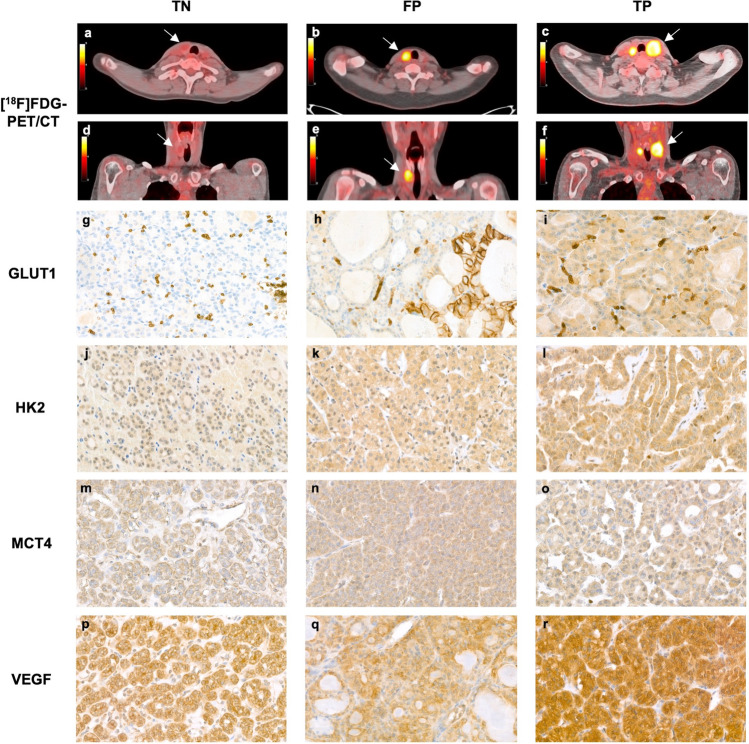
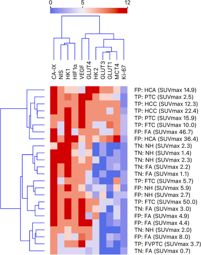
Procedures: Using a case-control design, 24 patients were selected from participants of a randomized controlled multicenter trial (NCT02208544) in which [18F]FDG-PET/CT and thyroid surgery were performed for Bethesda III and IV nodules. Three equally sized groups of [18F]FDG-positive malignant, [18F]FDG-positive benign, and [18F]FDG-negative benign nodules were included. Immunohistochemical staining was performed for glucose transporters (GLUT) 1, 3, and 4; hexokinases (HK) 1 and 2; hypoxia-inducible factor-1 alpha (HIF1α; monocarboxylate transporter 4 (MCT4); carbonic anhydrase IX (CA-IX); vascular endothelial growth factor (VEGF); sodium-iodide symporter (NIS); and Ki-67. Marker expression was scored using an immunoreactive score. Unsupervised cluster analysis was performed. The immunoreactive score was correlated to the maximum and peak standardized uptake values (SUVmax, SUVpeak) and SUVmax ratio (SUVmax of nodule/background SUVmax of contralateral, normal thyroid) of the [18F]FDG-PET/CT using the Spearman's rank correlation coefficient and compared between the three groups using Kruskal-Wallis tests.

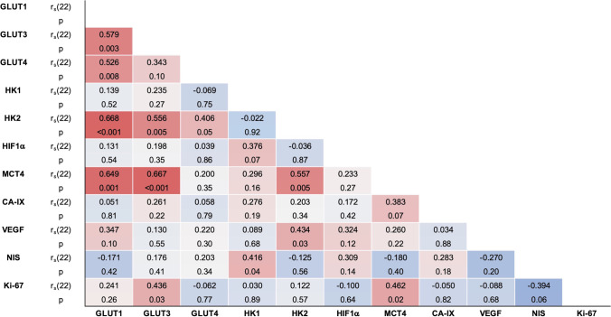
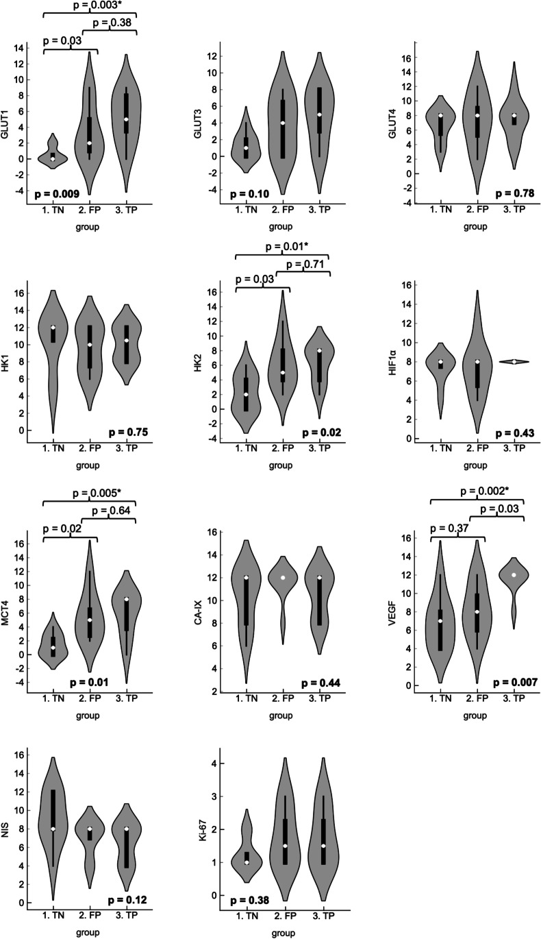
Results: The expression of GLUT1, GLUT3, HK2, and MCT4 was strongly positively correlated with the SUVmax, SUVpeak, and SUVmax ratio. The expression of GLUT1 (p = 0.009), HK2 (p = 0.02), MCT4 (p = 0.01), and VEGF (p = 0.007) was statistically significantly different between [18F]FDG-positive benign nodules, [18F]FDG-positive thyroid carcinomas, and [18F]FDG-negative benign nodules. In both [18F]FDG-positive benign nodules and [18F]FDG-positive thyroid carcinomas, the expression of GLUT1, HK2, and MCT4 was increased as compared to [18F]FDG-negative benign nodules. VEGF expression was higher in [18F]FDG-positive thyroid carcinomas as compared to [18F]FDG-negative and [18F]FDG-positive benign nodules.

Conclusions: Our results suggest that [18F]FDG-positive benign thyroid nodules undergo changes in protein expression similar to those in thyroid carcinomas. To expand the understanding of the metabolic changes in benign and malignant thyroid nodules, further research is required, including correlation with underlying genetic alterations.

Keywords: Glucose Metabolism; Glycolysis; Immunohistochemistry; Thyroid Nodule; [18F]FDG-PET/CT.


Conflict of interest statement

The authors declare no competing interests.

Figures

Fig. 1
Fig. 1
Representative [18F]FDG-PET/CT images and immunohistochemical staining patterns for the three groups. Transverse and coronal [18F]FDG-PET/CT images of a right-sided, 19-mm, visually [18F]FDG-negative follicular adenoma (TN group) with a SUVmax of 2.2 g/mL (a, d), a right-sided, 25-mm, visually [18F]FDG-positive follicular adenoma (FP group) with a SUVmax of 8.0 g/mL (b, e), and a left-sided, 40-mm, visually [18F]FDG-positive minimally invasive FTC (pT2N0Mx, TP group) with a SUVmax of 10.0 g/mL (with contralateral [18F]FDG-positive multinodular goiter) (c, f). Illustrative, representative microscopy images (× 40) of the immunohistochemistry stains observed in each of the groups show absent GLUT1 (g), weak HK2 (j), weak MCT4 (m), and intermediate VEGF (p) expression in TN nodules; weak cytoplasmic GLUT1 (h), intermediate HK2 (k), intermediate MCT4 (n), and intermediate VEGF (q) expression in FP nodules; and intermediate GLUT1 (i), intermediate HK2 (l), intermediate MCT4 (o), and strong VEGF (r) expression in TP nodules. In addition, several FP and TP nodules showed strong membranous GLUT1 expression in < 10% of cells (h). Note: the [18F]FDG-PET/CT images and microscopy images in each column represent multiple patients from each group to represent the average findings per group; single patients with immunohistochemistry results consistent with the group average were not available. FP, false positives. FTC, follicular thyroid carcinoma. GLUT, glucose transporter. HK, hexokinase. MCT4, Monocarboxylate transporter 4. SUVmax, maximum standardized uptake value. TN, true negatives. TP, true positives. VEGF, vascular endothelial growth factor.
Fig. 2
Fig. 2
Dendrogram heatmap showing the unsupervised cluster analysis of all 11 immunohistochemical stains of the 24 thyroid nodules, including the group, histopathological diagnosis, and SUVmax of these nodules. The IRS is presented on a scale from 0 (dark blue, absent stain) to 12 (dark red, strong stain in < 80% of tumor cells). CA-IX, carbonic anhydrase IX. FA, follicular adenoma. FP, false positives. FTC, follicular thyroid carcinoma. FVPTC, follicular variant PTC. GLUT, glucose transporter. HCA, Hürthle cell adenoma. HCC, Hürthle cell carcinoma. HIF1α, Hypoxia-inducible factor-1 alpha. HK, hexokinase. IRS, immunoreactive score. MCT4, Monocarboxylate transporter 4. NH, nodular hyperplasia. NIS, sodium-iodide symporter. PTC, papillary thyroid carcinoma. SUVmax, maximum standardized uptake value. TN, true negatives. TP, true positives. VEGF, vascular endothelial growth factor.
Fig. 3
Fig. 3
Spearman’s rank correlation coefficients (rs, df = 22) of the IRS of the 11 immunohistochemical markers, presented on a color scale from − 1 (dark blue) to 1 (dark red). CA-IX, carbonic anhydrase IX. GLUT, glucose transporter. HIF1a, Hypoxia-inducible factor-1 alpha. HK, hexokinase. IRS, immunoreactive score. MCT4, Monocarboxylate transporter 4. NIS, sodium-iodide symporter. VEGF, vascular endothelial growth factor.
Fig. 4
Fig. 4
Violin plots demonstrating the between-group comparison of the IRS of all IHC stains, including the median (white dot), interquartile range (thick black whisker), range (thin black whisker), and density based on counts (kernel). The boldfaced p value (bottom of each plot) represents the overall p value (Kruskal–Wallis test). The horizontal braces and corresponding p values represent the p values between two groups (post hoc Dunn’s test). Single asterisk (*) indicates statistical significance after Bonferroni correction for multiple comparisons. CA-IX, carbonic anhydrase IX. FP, false positives. GLUT, glucose transporter. HIF1a, Hypoxia-inducible factor-1 alpha. HK, hexokinase. IRS, immunoreactive score. MCT4, Monocarboxylate transporter 4. NIS, sodium-iodide symporter. TN, true negatives. TP, true positives. VEGF, vascular endothelial growth factor.

References

    1. Boellaard R, Delgado-Bolton R, Oyen WJ, et al. FDG PET/CT: EANM procedure guidelines for tumour imaging: version 2.0. Eur J Nucl Med Mol Imaging. 2015;42:328–354. doi: 10.1007/s00259-014-2961-x. - DOI - PMC - PubMed
    1. Liberti MV, Locasale JW. The Warburg effect: how does it benefit cancer cells? Trends Biochem Sci. 2016;41:211–218. doi: 10.1016/j.tibs.2015.12.001. - DOI - PMC - PubMed
    1. Piccardo A, Puntoni M, Dezzana M, et al. Indeterminate thyroid nodules. The role of (18)F-FDG PET/CT in the “era” of ultrasonography risk stratification systems and new thyroid cytology classifications. Endocrine. 2020;69:553–561. doi: 10.1007/s12020-020-02239-y. - DOI - PubMed
    1. Piccardo A, Puntoni M, Bertagna F, et al. 18F-FDG uptake as a prognostic variable in primary differentiated thyroid cancer incidentally detected by PET/CT: a multicentre study. Eur J Nucl Med Mol Imaging. 2014;41:1482–1491. doi: 10.1007/s00259-014-2774-y. - DOI - PubMed
    1. Santhanam P, Khthir R, Solnes LB, Ladenson PW. The relationship of BRAF(V600e) mutation status to FDG PET/CT avidity in thyroid cancer: a review and meta-analysis. Endocr Pract. 2018;24:21–26. doi: 10.4158/EP-2017-0080. - DOI - PubMed

Publication types

MeSH terms

Substances