Review
. 2022 Oct 17;17(1):83.
doi: 10.1186/s13000-022-01258-9.

A case of death of patient with ovarian fibroma combined with Meigs Syndrome and literature review

Affiliations
Review

A case of death of patient with ovarian fibroma combined with Meigs Syndrome and literature review

Na Tan et al. Diagn Pathol. .

Abstract

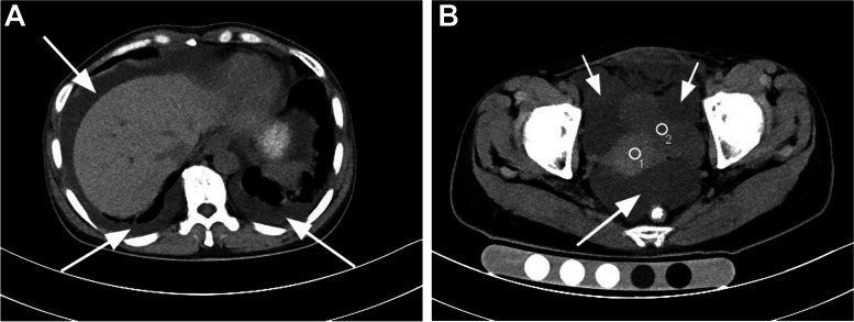
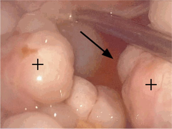
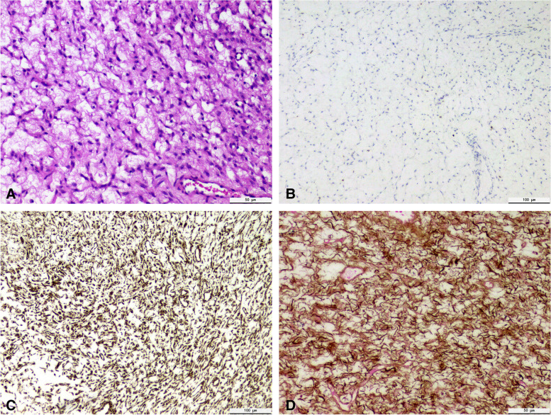
Ovarian fibroma is the most common benign pure stromal tumor. It has no specific clinical manifestation, most of which are pelvic or adnexal masses. 10-15% of cases with hydrothorax or ascites, after tumor resection, hydrothorax and ascites disappear, known as Meigs Syndrome. The elevated level of CA125 in a few patients was easily misdiagnosed as ovarian malignant tumor. A case of bilateral Ovarian fibroma associated with Meigs Syndrome is reported and the literature is reviewed in order to improve the understanding of the changes and avoid misdiagnosis.

Keywords: Meigs Syndrome; Ovarian fibroma.


Conflict of interest statement

The authors declare that they have no competing interests.

Figures

Fig. 1
Fig. 1
A and B, CT coronal images through the 10th thoracic vertebrae and the pelvis demonstrate there were multiple irregular cystic and solid masses in bilateral adnexa (White small arrow). A large amount of effusion (white big arrowhead) is present within the bilateral thoracic cavity and pelvic and abdominal cavity. the uterus (circle 1), the bladder (circle 2)
Fig. 2
Fig. 2
Laparoscopic Intraoperative photograph, can be found bilateral ovarian fibroma (black cross) and ascites (black arrow)
Fig. 3
Fig. 3
A Histology: showed a large number of spindle cells or ovoid cells, Tumor cell nucleus is long fusiform. There is an obvious edema between cells (400×). B Immunohistochemistry: α-inhibin weakly positive (100×). C Immunohistochemistry: Vimentin positive (100×). D Immunohistochemistry: Reticular fiber positive (400×)

References

    1. Chung BM, Park SB, Lee JB, Park HJ, Kim YS, Oh YJ. Magnetic resonance imaging features of ovarian fibroma, fibrothecoma, and thecoma. Abdom Imaging. 2015;40(5):1263–72. doi: 10.1007/s00261-014-0257-z. - DOI - PubMed
    1. Chen YJ, Hsieh CS, Eng HL, Huang CC. Ovarian fibroma in a 7-month-old infant: a case report and review of the literature. Pediatr Surg Int. 2004;20(11–12):894–7. doi: 10.1007/s00383-004-1284-6. - DOI - PubMed
    1. Zhang Z, Wu Y, Gao J. CT diagnosis in the thecoma-fibroma group of the ovarian stromal tumors. Cell Biochem Biophys. 2015;71(2):937–43. doi: 10.1007/s12013-014-0288-7. - DOI - PubMed
    1. Hogan ME, Andreotti R. Ovarian Fibroma Presenting With Meigs Syndrome. Ultrasound Q. 2015;31(3):215–8. doi: 10.1097/RUQ.0000000000000144. - DOI - PubMed
    1. Numanoglu C, Kuru O, Sakinci M, Akbayır O, Ulker V. Ovarian fibroma/fibrothecoma: retrospective cohort study shows limited value of risk of malignancy index score. Aust N Z J Obstet Gynaecol. 2013;53(3):287–92. doi: 10.1111/ajo.12090. - DOI - PubMed

Supplementary concepts